Femur
The femur, the thigh bone, is the only bone of the thigh. No other bone in the body is as long or as strong. (OpenStax A&P 2e, 8.4; StatPearls, Femur; Gray’s Anatomy (1918), Femur, p. 242)
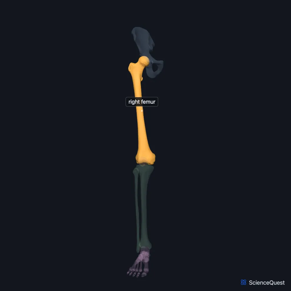
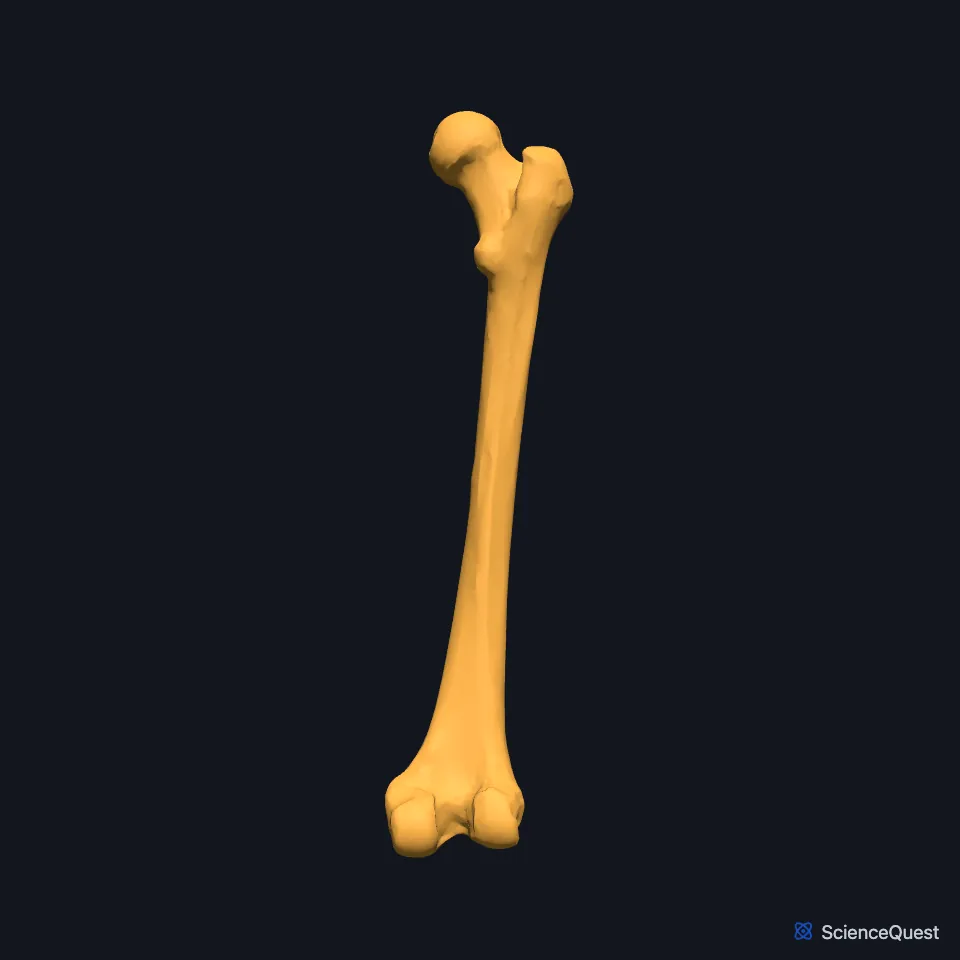
Open it in the Lower Limb Skeleton Explorer See it alone in 3D
Where is the femur?
The femur lies in the thigh, between the hip joint and the knee joint. The hips are wider than the knees, so the femur runs on a diagonal, while the tibia below it is close to vertical. Gray’s Anatomy (1918) calls the tibia vertical in males and slightly oblique in females. (OpenStax A&P 2e, 8.4; Gray’s Anatomy (1918), Femur, p. 242 and Tibia, p. 256; StatPearls, Femur)
You can feel its greater trochanter, which projects a little outward and backward where the neck meets the shaft. (OpenStax A&P 2e, 8.4; StatPearls, Hip Fracture Overview; Gray’s Anatomy (1918), Femur, p. 242)
What are the parts of the femur?
The rounded head at the top meets the acetabulum of the hip bone. A small pit on it, the fovea capitis, anchors the ligament of the head of the femur, which carries an artery to the head. (OpenStax A&P 2e, 8.4; StatPearls, Femur and Hip Joint; Gray’s Anatomy (1918), Femur, p. 242)
Below the head, the neck is a common place for the femur to break. The greater trochanter rises above the base of the neck, and the smaller lesser trochanter lies on the medial side just below it. (OpenStax A&P 2e, 8.4; StatPearls, Hip Fracture Overview; Gray’s Anatomy (1918), Femur, p. 242)
A broken hip, common in older people whose bones osteoporosis has weakened, is a fracture of the femoral neck. (OpenStax A&P 2e, 9.6; StatPearls, Hip Fracture Overview)
The neck meets the shaft at about 128 degrees in the average adult, and the angle decreases with age. (StatPearls, Femur)
The shaft bows forward a little. Down its back runs the linea aspera, a rough ridge where many hip and thigh muscles make long, thin attachments. (OpenStax A&P 2e, 8.4; Gray’s Anatomy (1918), Femur, p. 242; StatPearls, Femur)
At the lower end, the medial and lateral condyles meet the tibia. In front, their smooth surfaces join in the patellar surface, a groove for the patella. (OpenStax A&P 2e, 8.4; StatPearls, Femur; Gray’s Anatomy (1918), Femur, p. 242)
What does the femur do?
The femur bears weight and keeps the gait stable: the weight of the upper body rests on the two femoral heads. (StatPearls, Femur)
Muscles that act across the hip attach to the greater trochanter, and because it projects from the bone it gives them extra leverage. (OpenStax A&P 2e, 8.4; Gray’s Anatomy (1918), Femur, p. 242; StatPearls, Femur)
Which joints does the femur form, and what supplies its head?
At the hip, three ligaments from the hip bone spiral around the head and neck of the femur. Standing upright extends the hip and tightens them, and Gray’s credits the iliofemoral ligament with holding the body erect without muscular fatigue. (OpenStax A&P 2e, 9.6; StatPearls, Hip and Hip Joint; Gray’s Anatomy (1918), Hip joint, p. 333)
At the knee, each condyle meets a meniscus and the matching condyle of the tibia, and the patella forms a third joint with the groove at the front. (OpenStax A&P 2e, 9.6; Gray’s Anatomy (1918), Knee joint, p. 339; StatPearls, Knee)
The medial and lateral circumflex femoral arteries, usually branches of the deep femoral artery, supply the femoral head together with the obturator artery. (StatPearls, Femur and Femoral Artery)
How does the angle of the femur differ between people?
The femur slants inward from hip to knee, because the hips are set wider apart than the knees. The slant is greater in females, whose pelvis is wider. (OpenStax A&P 2e, 8.4; Gray’s Anatomy (1918), Femur, p. 242)
Where the sources differ
The sources define the Q-angle differently. OpenStax calls it how far the femur is angled laterally away from vertical, while StatPearls measures it between a line from the anterior superior iliac spine to the center of the patella and a line from there to the tibial tubercle. Both note a larger average Q-angle in females. (OpenStax A&P 2e, 8.4; StatPearls, Patellar Instability and Lateral Patellar Compression Syndrome)
| Feature | Detail and source |
|---|---|
| Bone type | Long bone, the longest and strongest in the body (OpenStax A&P 2e, 6.2 and 8.4; StatPearls, Femur; Gray’s Anatomy (1918), Classes of bones, p. 79) |
| Articulates with | Hip bone (hip joint); tibia (knee joint); patella (patellar surface) (OpenStax A&P 2e, 8.4; StatPearls, Femur and Hip Joint; Gray’s Anatomy (1918), Patella, p. 255) |
| Neck to shaft angle | About 128 degrees in the average adult (StatPearls, Femur) |
Touches in this model
Measured on the BodyParts3D meshes: surfaces within 2 mm of the femur, nearest first, 12 of 31.
- Lateral head of right gastrocnemius in contact
- Medial head of right gastrocnemius in contact
- Right adductor brevis in contact
- Right adductor longus in contact
- Right adductor magnus in contact
- Right adductor minimus in contact
- Right gemellus inferior in contact
- Right gemellus superior in contact
- Right gluteus maximus in contact
- Right gluteus medius in contact
- Right gluteus minimus in contact
- Right iliacus in contact
Sources
- OpenStax Anatomy and Physiology 2e, section 8.4, Bones of the Lower Limb. CC BY-NC-SA 4.0. Read 2026-10-25.
- StatPearls, Anatomy, Bony Pelvis and Lower Limb: Femur. Amy Chang; Grant Breeland; Asa C. Black; John B. Hubbard. Last update November 17, 2023. CC BY-NC-ND 4.0. Read 2026-10-25.
- Gray’s Anatomy of the Human Body, 20th US edition (1918), edited by Warren H. Lewis, Osteology, The Femur (pp. 242 to 255). Henry Gray; Warren H. Lewis (editor). Public domain. Read 2026-10-25.
- Gray’s Anatomy of the Human Body, 20th US edition (1918), edited by Warren H. Lewis, Osteology, The Tibia (pp. 256 to 260); 1918 name: intercondyloid eminence for intercondylar eminence. Henry Gray; Warren H. Lewis (editor). Public domain. Read 2026-10-25.
- StatPearls, Hip Fracture Overview. Benjamin R. Emmerson; Matthew A. Varacallo; Dominic Inman. Last update August 8, 2023. CC BY-NC-ND 4.0. Read 2026-10-25.
- StatPearls, Anatomy, Bony Pelvis and Lower Limb, Hip Joint. Maks Gold; Akul Munjal; Matthew A. Varacallo. Last update July 25, 2023. CC BY-NC-ND 4.0. Read 2026-10-25.
- OpenStax Anatomy and Physiology 2e, section 9.6, Anatomy of Selected Synovial Joints. CC BY-NC-SA 4.0. Read 2026-10-25.
- StatPearls, Anatomy, Bony Pelvis and Lower Limb, Hip. Roland Glenister; Sandeep Sharma. Last update July 24, 2023. CC BY-NC-ND 4.0. Read 2026-10-25.
- Gray’s Anatomy of the Human Body, 20th US edition (1918), edited by Warren H. Lewis, Syndesmology, Coxal Articulation or Hip-joint (pp. 333 to 339). Henry Gray; Warren H. Lewis (editor). Public domain. Read 2026-10-25.
- Gray’s Anatomy of the Human Body, 20th US edition (1918), edited by Warren H. Lewis, Syndesmology, The Knee-joint (pp. 339 to 347). Henry Gray; Warren H. Lewis (editor). Public domain. Read 2026-10-25.
- StatPearls, Anatomy, Bony Pelvis and Lower Limb, Knee. Marco Gupton; Onyebuchi Imonugo; Asa C. Black; Marjorie V. Launico; Robert R. Terreberry. Last update November 5, 2023. CC BY-NC-ND 4.0. Read 2026-10-25.
- StatPearls, Anatomy, Bony Pelvis and Lower Limb: Femoral Artery. Hilary Swift; Bruno Bordoni. Last update November 29, 2023. CC BY-NC-ND 4.0. Read 2026-10-25.
- OpenStax Anatomy and Physiology 2e, section 6.2, Bone Classification. CC BY-NC-SA 4.0. Read 2026-10-25.
- Gray’s Anatomy of the Human Body, 20th US edition (1918), edited by Warren H. Lewis, Osteology, introduction: long, short, flat and irregular bones (p. 79). Henry Gray; Warren H. Lewis (editor). Public domain. Read 2026-10-25.
- Gray’s Anatomy of the Human Body, 20th US edition (1918), edited by Warren H. Lewis, Osteology, The Patella (pp. 255 to 256). Henry Gray; Warren H. Lewis (editor). Public domain. Read 2026-10-25.
- StatPearls, Patellar Instability. Steve Wolfe; Matthew A. Varacallo; Joshua D. Thomas; Jeffrey J. Carroll; Chadi I. Kahwaji. Last update September 4, 2023. CC BY-NC-ND 4.0. Read 2026-10-25.
- StatPearls, Lateral Patellar Compression Syndrome. Amin Abukar; Daniel Li. Last update August 2, 2025. CC BY-NC-ND 4.0. Read 2026-10-25.
The values are facts and free to use; this page’s selection and presentation are © 2026 ScienceQuest.