Iliacus
The iliacus is a fan-shaped muscle whose back surface rests in the iliac fossa of the pelvis. With the psoas major it makes up the iliopsoas, the primary flexor of the thigh. (StatPearls, Iliopsoas Muscle, Psoas Major and Anterior Thigh Muscles; OpenStax A&P 2e, 11.6; Gray’s Anatomy (1918), Iliac Region)
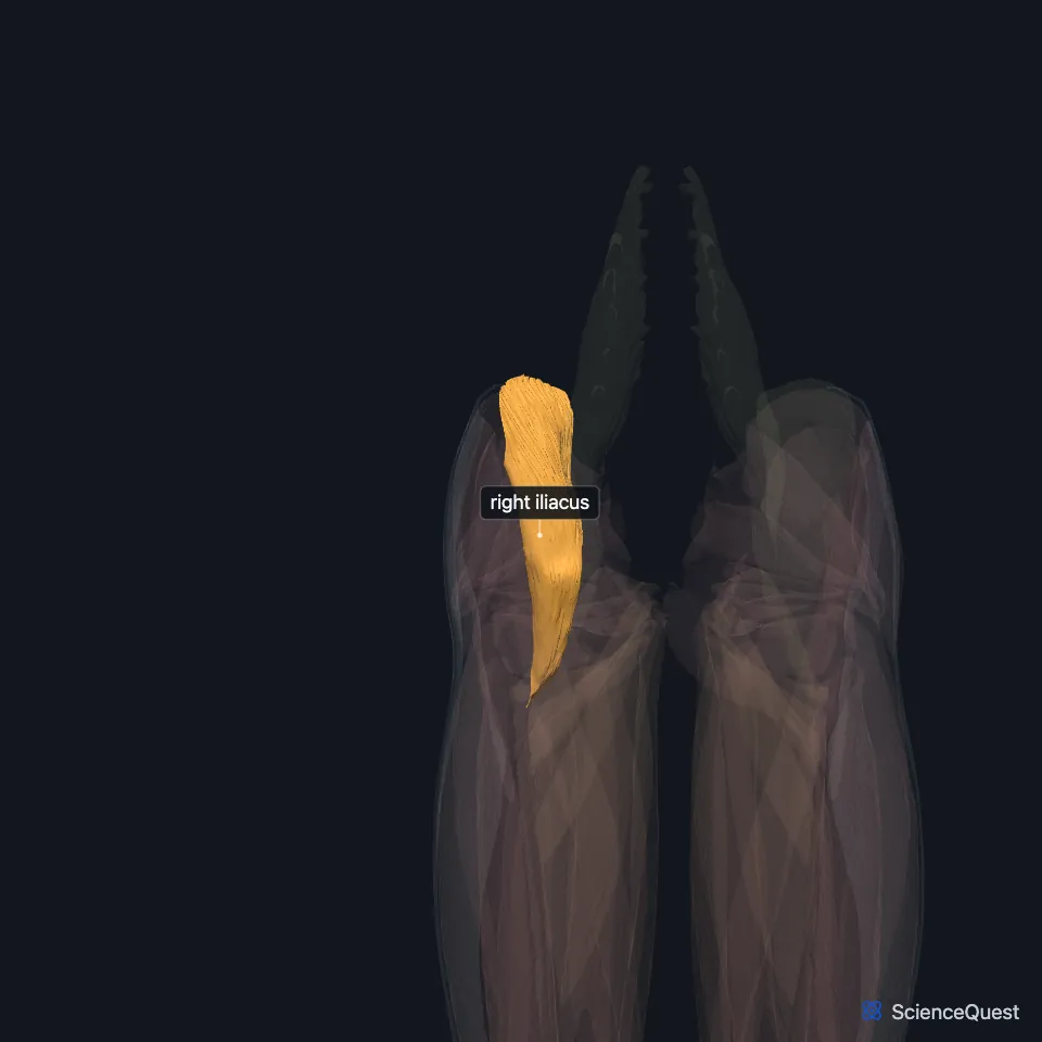
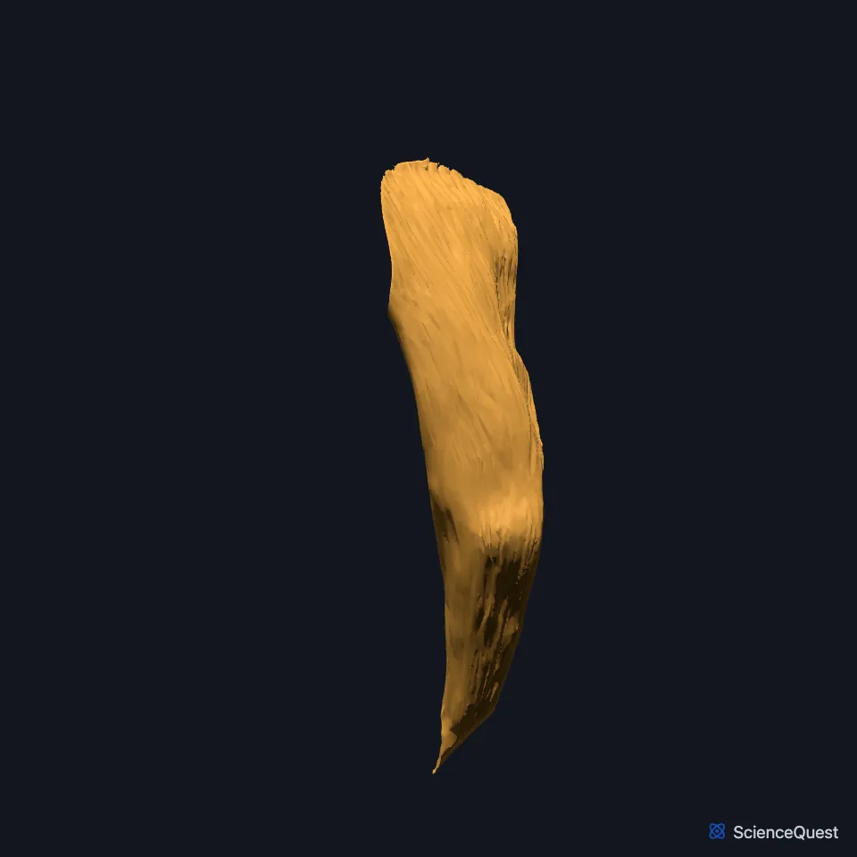
Open it in the Lower Limb Muscles Explorer See it alone in 3D
Where is the iliacus?
It arises from the upper two-thirds of the iliac fossa and the side of the sacral wing. Its fibers pass under the inguinal ligament, in front of the hip joint, with those of psoas major. (StatPearls, Iliopsoas Muscle)
Inside the pelvis it lies behind the cecum on the right and the sigmoid colon on the left. (StatPearls, Psoas Major)
Where does the iliacus insert?
Its bundles merge into the tendon of psoas major and insert with it on the lesser trochanter of the femur. A few lateral and lower bundles attach to the femur directly. (StatPearls, Iliopsoas Muscle and Psoas Major)
The iliopectineal bursa lies under the shared tendon and separates it from the lesser trochanter and the upper femur. (StatPearls, Iliopsoas Muscle)
What does the iliacus do?
As part of the iliopsoas it flexes the thigh and adds to adduction and lateral rotation at the hip. With the lower limb fixed, the iliopsoas flexes the trunk and bends it to the same side. (StatPearls, Iliopsoas Muscle)
It can also act alone, steadying the pelvis and helping to flex the hip during running. (StatPearls, Iliopsoas Muscle)
What lies next to the iliacus?
The femoral nerve supplies it. On its way to the thigh the nerve runs between the iliacus and psoas major. (StatPearls, Iliopsoas Muscle and Thigh Femoral Nerve)
In the thigh, the iliopsoas forms part of the floor of the femoral triangle and rests on the fibrous capsule of the hip joint. Sartorius and rectus femoris run along its outer edge. (StatPearls, Adductor Canal and Psoas Major)
The external iliac artery supplies it through several muscular branches. (StatPearls, Iliopsoas Muscle)
Where the sources differ
Sources differ on the spinal roots behind its supply. The StatPearls anterior thigh article gives the femoral nerve to the iliacus as L1 to L4, and its thigh muscles article gives the muscular branch as L1 to L3. Its iliopsoas article gives the femoral nerve to the iliacus as L2 to L4, and its femoral nerve article has the nerve forming from L2 to L4. (StatPearls, Anterior Thigh Muscles, Thigh Muscles, Iliopsoas Muscle and Thigh Femoral Nerve)
Sources differ on whether the iliopsoas adducts or abducts the femur. The StatPearls iliopsoas article says the unit adds to adduction. Its psoas major article says in one passage that contraction abducts the femur, and in its function section that the psoas complex adducts it. (StatPearls, Iliopsoas Muscle and Psoas Major)
| Feature | Detail and source |
|---|---|
| Origin | Upper two-thirds of the iliac fossa; side of the sacral wing (StatPearls, Iliopsoas Muscle) |
| Insertion | Lesser trochanter of the femur, through the psoas major tendon (StatPearls, Iliopsoas Muscle) |
| Action | Flexes the thigh at the hip, as part of the iliopsoas; steadies the pelvis (StatPearls, Iliopsoas Muscle) |
| Nerve | Femoral nerve (StatPearls, Iliopsoas Muscle) |
Touches in this model
Measured on the BodyParts3D meshes: surfaces within 2 mm of the iliacus, nearest first, 12 of 14.
- Right psoas major in contact
- Right rectus femoris in contact
- Right sartorius in contact
- Colon in contact
- Ileum in contact
- Omental taenia in contact
- Right deferent duct in contact
- Right femur in contact
- Right hip bone in contact
- Right transversus abdominis in contact
- Skin in contact
- Right inguinal ligament 0.8 mm away
Sources
- StatPearls, Anatomy, Bony Pelvis and Lower Limb: Iliopsoas Muscle. Bruno Bordoni; Matthew A. Varacallo. Last update June 8, 2026. CC BY-NC-ND 4.0. Read 2026-10-25.
- StatPearls, Anatomy, Bony Pelvis and Lower Limb: Psoas Major. Marco A. Siccardi; Muhammad Ali Tariq; Cristina Valle. Last update August 8, 2023. CC BY-NC-ND 4.0. Read 2026-10-25.
- StatPearls, Anatomy, Bony Pelvis and Lower Limb: Anterior Thigh Muscles. Ayesha Khan; Abdul Arain. Last update March 17, 2023. CC BY-NC-ND 4.0. Read 2026-10-25.
- OpenStax Anatomy and Physiology 2e, section 11.6, Appendicular Muscles of the Pelvic Girdle and Lower Limbs. CC BY-NC-SA 4.0. Read 2026-10-25.
- Anatomy of the Human Body (Gray’s Anatomy), 20th US edition, 1918, Myology, The Muscles and Fasciae of the Iliac Region, pages 466 to 467, with the footnote on the Iliopsoas on page 467; Internet Archive scan of the 1918 Lea and Febiger printing, terminology of 1918. Henry Gray; edited by Warren H. Lewis. Public domain. Read 2026-10-25.
- StatPearls, Anatomy, Bony Pelvis and Lower Limb: Thigh Femoral Nerve. Nader A. Refai; Asa C. Black; Prasanna Tadi. Last update August 22, 2023. CC BY-NC-ND 4.0. Read 2026-10-25.
- StatPearls, Anatomy, Abdomen and Pelvis: Adductor Canal (Subsartorial Canal, Hunter Canal). Allan Migirov; Tafline C. Arbor; Renato C. Vilella. Last update January 9, 2024. CC BY-NC-ND 4.0. Read 2026-10-25.
- StatPearls, Anatomy, Bony Pelvis and Lower Limb: Thigh Muscles. Basem Attum; Matthew A. Varacallo. Last update December 17, 2022. CC BY-NC-ND 4.0. Read 2026-10-25.
The values are facts and free to use; this page’s selection and presentation are © 2026 ScienceQuest.