Ileum
The ileum is the final and longest part of the small intestine. The ileocecal valve separates it from the cecum, the first part of the large intestine. (OpenStax A&P 2e, 23.5; StatPearls, Physiology, Small Bowel, Small Intestine and Large Intestine)
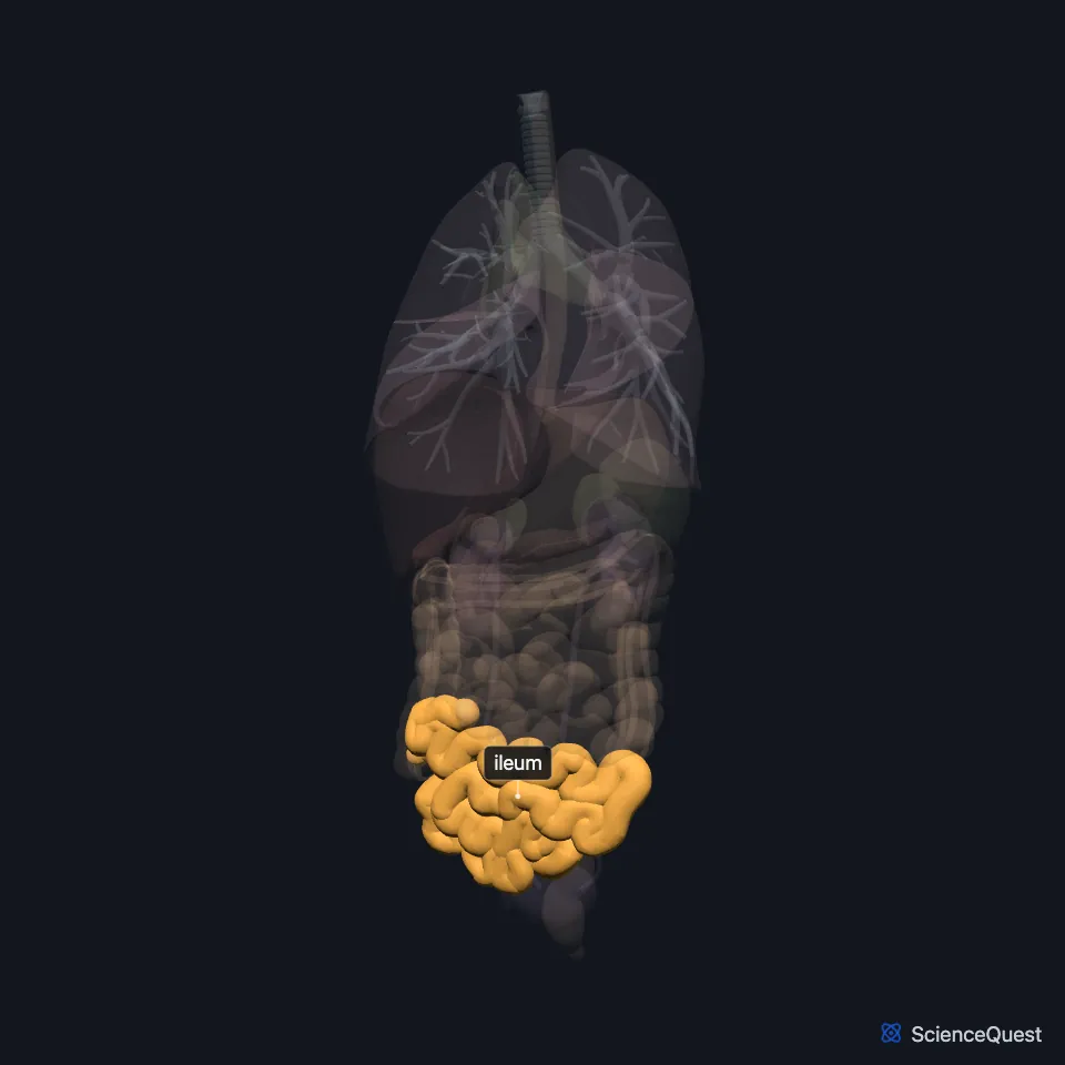
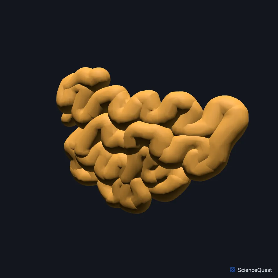
Where is the ileum?
It follows the jejunum with no clear boundary and opens into the cecum through its medial wall, in the right iliac fossa. (OpenStax A&P 2e, 23.5; StatPearls, Large Intestine)
Like the jejunum, it hangs from the posterior abdominal wall on the mesentery, framed by the colon and the rest of the large intestine. (OpenStax A&P 2e, 23.5; StatPearls, Small Intestine and Physiology, Small Bowel)
How is the ileum built?
Clusters of lymphoid follicles called Peyer’s patches are densest in the distal ileum. The small intestine also acts as an immune barrier to gut bacteria. (OpenStax A&P 2e, 23.5; StatPearls, Small Intestine)
The circular folds of the small intestine grow fewer and smaller along the ileum and disappear toward its lower end. (OpenStax A&P 2e, 23.5; Gray’s Anatomy (1918), The Small Intestine)
Segmentation, the mixing contraction that churns the jejunum, slows in the ileum, where peristalsis takes over. (OpenStax A&P 2e, 23.5; StatPearls, Physiology, Small Bowel)
What does the ileum do?
It takes up what the duodenum and jejunum left behind, above all vitamin B12 and bile acids. (StatPearls, Small Intestine)
Most bile salts are absorbed at the end of the ileum and carried in the blood back to the liver, which secretes them again. This loop is the enterohepatic circulation. (OpenStax A&P 2e, 23.6; StatPearls, Physiology, Bile Acids)
Vitamin B12 uptake in the terminal ileum depends on intrinsic factor, made by parietal cells in the stomach. (StatPearls, Stomach)
What supplies the ileum?
Ileal arteries from the superior mesenteric artery run in the mesentery and form arcades and vasa recta, as in the jejunum. Its veins drain to the superior mesenteric vein and on to the portal vein. (StatPearls, Small Intestine)
Its lymph drains to nodes in the mesentery and along the superior mesenteric artery, then to the cisterna chyli and the thoracic duct. This route is a major path for absorbed fat. (StatPearls, Small Intestine)
Which conditions involve the ileum?
A Meckel diverticulum, when present, often lies near the ileocecal valve. The “rule of 2s” puts it about 2 feet from the valve, in about 2% of people. (StatPearls, Small Intestine)
Crohn disease, a chronic inflammatory condition, affects the ileum more often than any other part of the gut. (StatPearls, Small Intestine)
Where the sources differ
The sources disagree on how the ileum compares with the jejunum. OpenStax says the ileum is thicker, more vascular and has more developed mucosal folds; StatPearls says the jejunum has the thicker wall and greater vascularity, and the ileum more mesenteric fat and more Peyer patches. (OpenStax A&P 2e, 23.5; StatPearls, Small Intestine)
Its length differs by source: about 1.8 m (6 ft) in OpenStax, around 3 m in StatPearls. (OpenStax A&P 2e, 23.5; StatPearls, Small Intestine)
| Feature | Detail and source |
|---|---|
| Ends | At the cecum, through the ileocecal valve (OpenStax A&P 2e, 23.5; StatPearls, Physiology, Small Bowel) |
| Lymphoid tissue | Peyer’s patches, concentrated in the distal ileum (OpenStax A&P 2e, 23.5; StatPearls, Small Intestine) |
| Absorbs | Vitamin B12 and bile acids, with what the jejunum left (StatPearls, Small Intestine) |
| Arteries | Ileal arteries from the superior mesenteric artery (StatPearls, Small Intestine) |
Touches in this model
Measured on the BodyParts3D meshes: surfaces within 2 mm of the ileum, nearest first, 12 of 19.
- Colon in contact
- Free taenia in contact
- Jejunum in contact
- Omental taenia in contact
- Urinary bladder in contact
- Left external iliac vein in contact
- Left iliacus in contact
- Left transversus abdominis in contact
- Right external iliac artery in contact
- Right external iliac vein in contact
- Right iliacus in contact
- Skin in contact
Sources
- OpenStax Anatomy and Physiology 2e, section 23.5, The Small and Large Intestines. CC BY-NC-SA 4.0. Read 2026-10-25.
- StatPearls, Physiology, Small Bowel. Elizabeth M. Fish; Karlie R. Shumway; Bracken Burns. Last update January 31, 2024. CC BY-NC-ND 4.0. Read 2026-10-25.
- StatPearls, Anatomy, Abdomen and Pelvis, Small Intestine. Jason T. Collins; Amanda Nguyen; Adekunle E. Omole; Madhu Badireddy. Last update February 18, 2025. CC BY-NC-ND 4.0. Read 2026-10-25.
- StatPearls, Anatomy, Abdomen and Pelvis: Large Intestine. Adekunle E. Omole; Pujyitha Mandiga; Preet Kahai; Stany Lobo. Last update April 6, 2025. CC BY-NC-ND 4.0. Read 2026-10-25.
- Gray’s Anatomy of the Human Body, 20th US edition (1918), Splanchnology, The Small Intestine, page 1171. Henry Gray; edited by Warren H. Lewis. Public domain. Read 2026-10-25.
- OpenStax Anatomy and Physiology 2e, section 23.6, Accessory Organs in Digestion: The Liver, Pancreas, and Gallbladder. CC BY-NC-SA 4.0. Read 2026-10-25.
- StatPearls, Physiology, Bile Acids. Isaac Chen; Sebastiano Cassaro. Last update May 1, 2023. CC BY-NC-ND 4.0. Read 2026-10-25.
- StatPearls, Anatomy, Abdomen and Pelvis: Stomach. Shazia R. Chaudhry; Maria Nataly P. Liman; Adekunle E. Omole; Diana C. Peterson. Last update July 17, 2024. CC BY-NC-ND 4.0. Read 2026-10-25.
The values are facts and free to use; this page’s selection and presentation are © 2026 ScienceQuest.