Urinary bladder
The urinary bladder is a hollow muscular reservoir in the pelvis. It collects urine from both ureters and empties it through the urethra. (StatPearls, Abdomen and Pelvis: Bladder; OpenStax A&P 2e, 25.2)
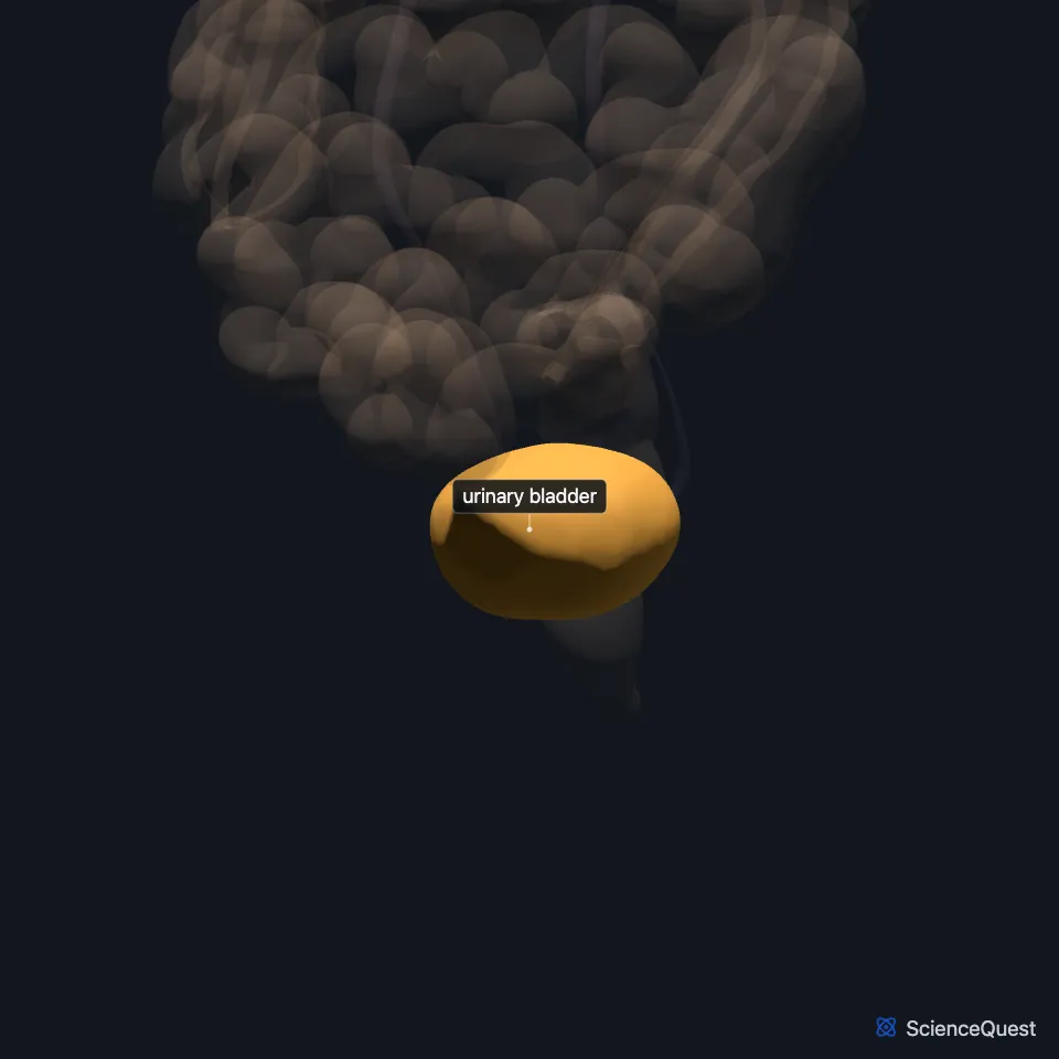
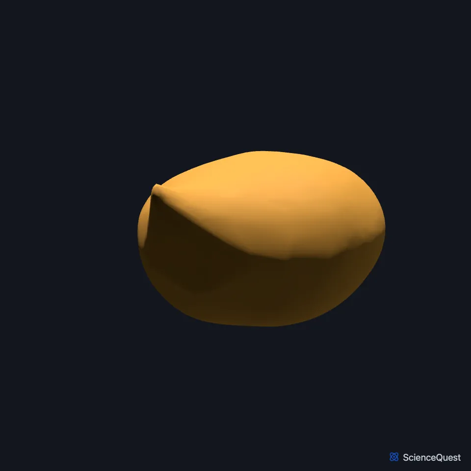
Where is the bladder?
When empty it lies in the lesser pelvis, just behind the pubic symphysis, and when full it rises into the abdominal cavity. In children it sits in the abdomen and does not fully descend into the pelvis until puberty. (StatPearls, Abdomen and Pelvis: Bladder)
What are the parts of the bladder?
It has four parts. The apex, or dome, points up toward the abdominal wall, the fundus, or base, lies at the back and bottom, the body lies between them, and the neck narrows into the urethra. (StatPearls, Abdomen and Pelvis: Bladder)
Its wall is mostly the detrusor, a smooth muscle. The lining is transitional epithelium, whose cells change shape and flatten as the bladder fills. (OpenStax A&P 2e, 25.2; StatPearls, Abdomen and Pelvis: Bladder; Gray 1918, 3b. 3. The Urinary Bladder)
The two ureteral openings and the start of the urethra mark the three corners of a triangle at the base of the bladder, the trigone. (OpenStax A&P 2e, 25.2; Gray 1918, 3b. 3. The Urinary Bladder)
How is the bladder controlled?
It stores urine until voiding, and the pressure inside rises as it fills, bringing first a sense of fullness and then pain. StatPearls gives 300 to 400 mL as the volume an adult bladder holds in comfort, while OpenStax puts the loss of voluntary control at that volume. (OpenStax A&P 2e, 25.2; StatPearls, Physiology, Bladder)
Parasympathetic nerves from S2 to S4 contract the detrusor and relax the internal urethral sphincter to empty it. Sympathetic nerves from T10 to L2 relax the detrusor and tighten the internal sphincter while it fills, and the pudendal nerve gives voluntary control of the external urethral sphincter. (StatPearls, Abdomen and Pelvis: Bladder)
Stretch receptors in the wall send signals to the central nervous system through the hypogastric and pelvic splanchnic nerves, and those signals change with how far the bladder is stretched. Signals from the upper wall travel with the sympathetic nerves, and those from the lower part with the parasympathetic nerves. (StatPearls, Abdomen and Pelvis: Bladder)
What lies next to the bladder?
In males the rectum lies behind the bladder and the prostate below it. In females the uterus and the front wall of the vagina lie behind it. (StatPearls, Abdomen and Pelvis: Bladder; OpenStax A&P 2e, 25.2)
The muscles of the pelvic diaphragm support it from below. Peritoneum covers its upper surface and part of its back, and endopelvic fascia covers its lower part and lower sides. (StatPearls, Abdomen and Pelvis: Bladder)
Most of its blood comes from the superior and inferior vesical arteries, indirect branches of the internal iliac arteries, with some from the obturator and inferior gluteal arteries. It drains through the vesical venous plexus to the internal iliac vein, and most of its lymph goes to the external iliac nodes. (StatPearls, Abdomen and Pelvis: Bladder)
Where the sources differ
OpenStax calls the bladder retroperitoneal, with peritoneum only over its dome. StatPearls calls it subperitoneal, with peritoneum over its upper surface and part of its back. (OpenStax A&P 2e, 25.2; StatPearls, Abdomen and Pelvis: Bladder)
OpenStax gives adult volumes from nearly zero to 500 to 600 mL. StatPearls says the bladder can usually hold up to 500 mL. (OpenStax A&P 2e, 25.2; StatPearls, Abdomen and Pelvis: Bladder)
OpenStax describes normal voiding as a spinal reflex through the sacral spinal cord. StatPearls says local spinal reflexes mainly run filling, but voiding needs the pontine micturition center in the pons. (OpenStax A&P 2e, 25.2; StatPearls, Abdomen and Pelvis: Bladder)
| Feature | Detail and source |
|---|---|
| Parts | Apex (dome), body, fundus (base), neck (StatPearls, Abdomen and Pelvis: Bladder) |
| Muscle | Detrusor, smooth muscle (OpenStax A&P 2e, 25.2; StatPearls, Abdomen and Pelvis: Bladder) |
| Arteries | Superior and inferior vesical arteries, with some from the obturator and inferior gluteal arteries (StatPearls, Abdomen and Pelvis: Bladder) |
| Nerves | Pelvic splanchnic (S2 to S4), hypogastric (T10 to L2), pudendal (S2 to S4) (StatPearls, Abdomen and Pelvis: Bladder) |
Touches in this model
Measured on the BodyParts3D meshes: surfaces within 2 mm of the urinary bladder, nearest first, 12 of 17.
- Ileum in contact
- Left ureter in contact
- Right ureter in contact
- Left deferent duct in contact
- Left iliococcygeus in contact
- Left pubococcygeus in contact
- Left puborectalis in contact
- Left seminal vesicle in contact
- Prostate in contact
- Right deferent duct in contact
- Right iliococcygeus in contact
- Right pubococcygeus in contact
Sources
- StatPearls, Anatomy, Abdomen and Pelvis: Bladder. Esra S. Shermadou; Sajedur Rahman; Stephen W. Leslie. Last update July 24, 2023. CC BY-NC-ND 4.0. Read 2026-10-25.
- OpenStax Anatomy and Physiology 2e, section 25.2, Gross Anatomy of Urine Transport. CC BY-NC-SA 4.0. Read 2026-10-25.
- Gray’s Anatomy of the Human Body, 20th US edition (1918), XI. Splanchnology, 3b. 3. The Urinary Bladder (1918 terms: trigonum vesicae for trigone). Henry Gray. Public domain. Read 2026-10-25.
- StatPearls, Physiology, Bladder. Nicholas J. Lanzotti; Muhammad Ali Tariq; Srinivasa Rao Bolla. Last update May 1, 2023. CC BY-NC-ND 4.0. Read 2026-10-25.
The values are facts and free to use; this page’s selection and presentation are © 2026 ScienceQuest.