Rectum
The rectum is the last part of the large intestine, running from the sigmoid colon to the anal canal. Its name comes from the Latin intestinum rectum, the straight intestine. (StatPearls, Rectum)
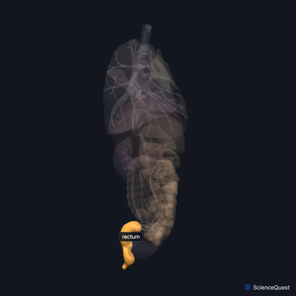
Where is the rectum?
Residue from the sigmoid colon enters it at the level of the third sacral vertebra. From there it follows the hollow of the sacrum. (OpenStax A&P 2e, 23.5; StatPearls, Rectum)
It is the most posterior organ in the pelvic cavity. In men the prostate lies in front of it; in women the cervix, uterus and vagina do. (StatPearls, Rectum)
It becomes the anal canal at the level of the levator ani muscle. (StatPearls, Rectum)
What shape is the rectum?
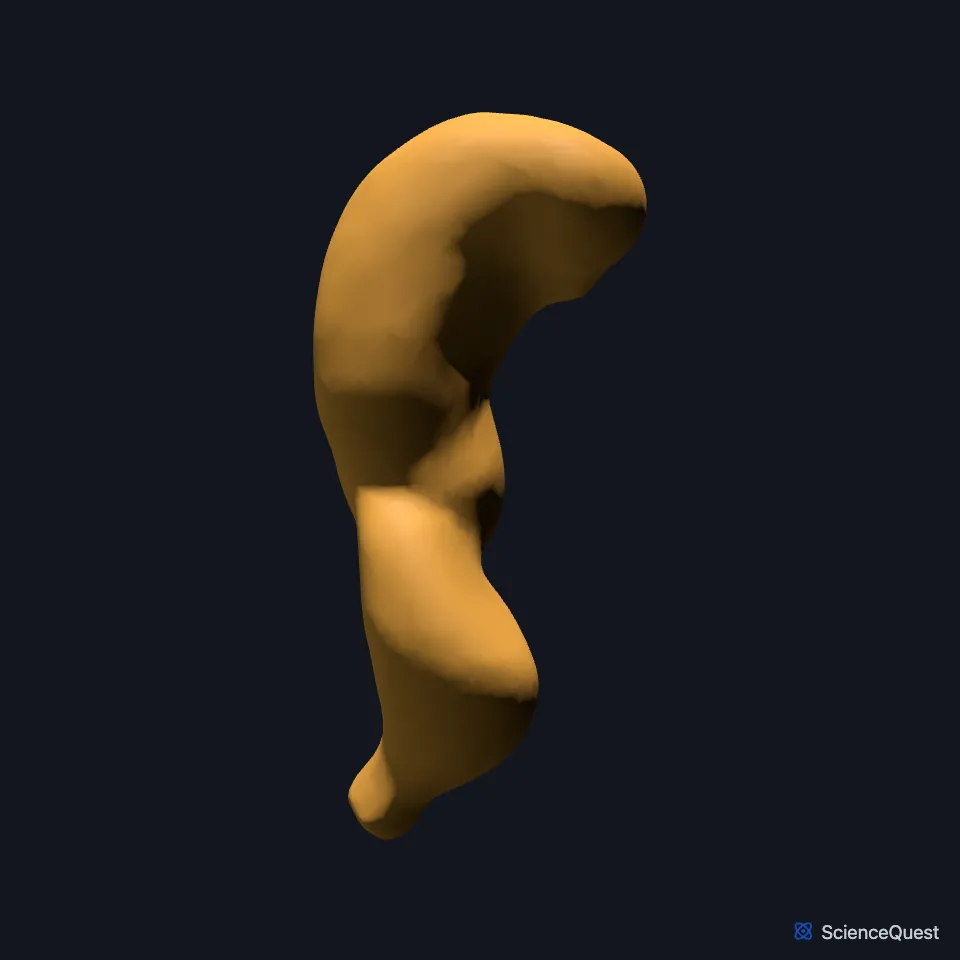
It curves twice from front to back: along the hollow of the sacrum at the sacral flexure, then forward at the anorectal flexure. Three lateral bends inside it form folds called the rectal valves, or valves of Houston. (StatPearls, Rectum; OpenStax A&P 2e, 23.5)
Where the colon ends, its three teniae coli spread out into a complete outer layer of longitudinal muscle. That is what sets the rectum apart from the colon, and it has no haustra. (StatPearls, Rectum; OpenStax A&P 2e, 23.5)
Peritoneum covers the front and sides of its upper third and only the front of its middle third. The lower third has none. (StatPearls, Rectum)
At the anorectal junction its inner circular muscle thickens into the internal anal sphincter. (StatPearls, Rectum)
What does the rectum do?
It stores stool until defecation and helps control defecation and continence. (StatPearls, Rectum)
You sense it filling through parasympathetic fibers that run to the S2, S3 and S4 spinal nerves. Its well-developed muscle layers produce the strong contractions of defecation. (StatPearls, Rectum; OpenStax A&P 2e, 23.5)
The pelvic floor muscles support it and help with defecation and continence. Most of the floor is the levator ani: the iliococcygeus, pubococcygeus and puborectalis. (StatPearls, Rectum)
What supplies the rectum?
The superior rectal artery, the end of the inferior mesenteric artery, supplies it. The middle rectal artery, a branch of the internal iliac artery, adds blood to its lower part. (StatPearls, Rectum)
Most of its blood drains through the superior rectal vein and the inferior mesenteric vein to the portal system. The lower part drains through the middle and inferior rectal veins to the internal iliac veins. (StatPearls, Rectum)
Sympathetic fibers come from the lumbar splanchnic nerves, roots L1 to L2. Parasympathetic fibers come from the sacral region and join them in the pelvic plexus. (StatPearls, Rectum)
In men the peritoneum folds from the rectum onto the back of the bladder, forming the rectovesical pouch. In women it folds onto the back of the cervix, forming the rectouterine pouch, or pouch of Douglas. (StatPearls, Rectum)
Where the sources differ
Its length depends on the source: OpenStax calls the rectum the final 20.3 cm (8 in) of the alimentary canal, while StatPearls gives 12 to 15 cm from the rectosigmoid junction to the dentate line. (OpenStax A&P 2e, 23.5; StatPearls, Rectum)
The two StatPearls articles place the ampulla, the widened reservoir of the rectum, differently: the rectum article calls it the final portion, resting on the pelvic diaphragm, and the large intestine article the middle segment. (StatPearls, Rectum and Large Intestine)
| Feature | Detail and source |
|---|---|
| Begins | Near the third sacral vertebra, continuing the sigmoid colon (OpenStax A&P 2e, 23.5; StatPearls, Rectum) |
| Ends | Anal canal, at the levator ani (StatPearls, Rectum) |
| Folds | Three rectal valves (valves of Houston) (OpenStax A&P 2e, 23.5; StatPearls, Rectum) |
| Arteries | Superior rectal (from the inferior mesenteric); middle rectal (from the internal iliac), to the lower rectum (StatPearls, Rectum) |
| Nerves | Lumbar splanchnic (L1 to L2) and sacral parasympathetic fibers, via the pelvic plexus (StatPearls, Rectum) |
Touches in this model
Measured on the BodyParts3D meshes: surfaces within 2 mm of the rectum, nearest first, 12 of 22.
- Colon in contact
- Free taenia in contact
- Mesocolic taenia in contact
- Omental taenia in contact
- External anal sphincter in contact
- Left deferent duct in contact
- Left iliococcygeus in contact
- Left pubococcygeus in contact
- Left puborectalis in contact
- Right deferent duct in contact
- Right iliococcygeus in contact
- Right pubococcygeus in contact
Sources
- StatPearls, Anatomy, Abdomen and Pelvis, Rectum. Yun Hwa W. Wang; Jeffrey Wiseman. Last update July 25, 2023. CC BY-NC-ND 4.0. Read 2026-10-25.
- OpenStax Anatomy and Physiology 2e, section 23.5, The Small and Large Intestines. CC BY-NC-SA 4.0. Read 2026-10-25.
- StatPearls, Anatomy, Abdomen and Pelvis: Large Intestine. Adekunle E. Omole; Pujyitha Mandiga; Preet Kahai; Stany Lobo. Last update April 6, 2025. CC BY-NC-ND 4.0. Read 2026-10-25.
The values are facts and free to use; this page’s selection and presentation are © 2026 ScienceQuest.