Colon
The colon is the part of the large intestine between the cecum and the rectum. It runs as the ascending, transverse, descending and sigmoid colon. (OpenStax A&P 2e, 23.5; StatPearls, Physiology, Large Intestine)
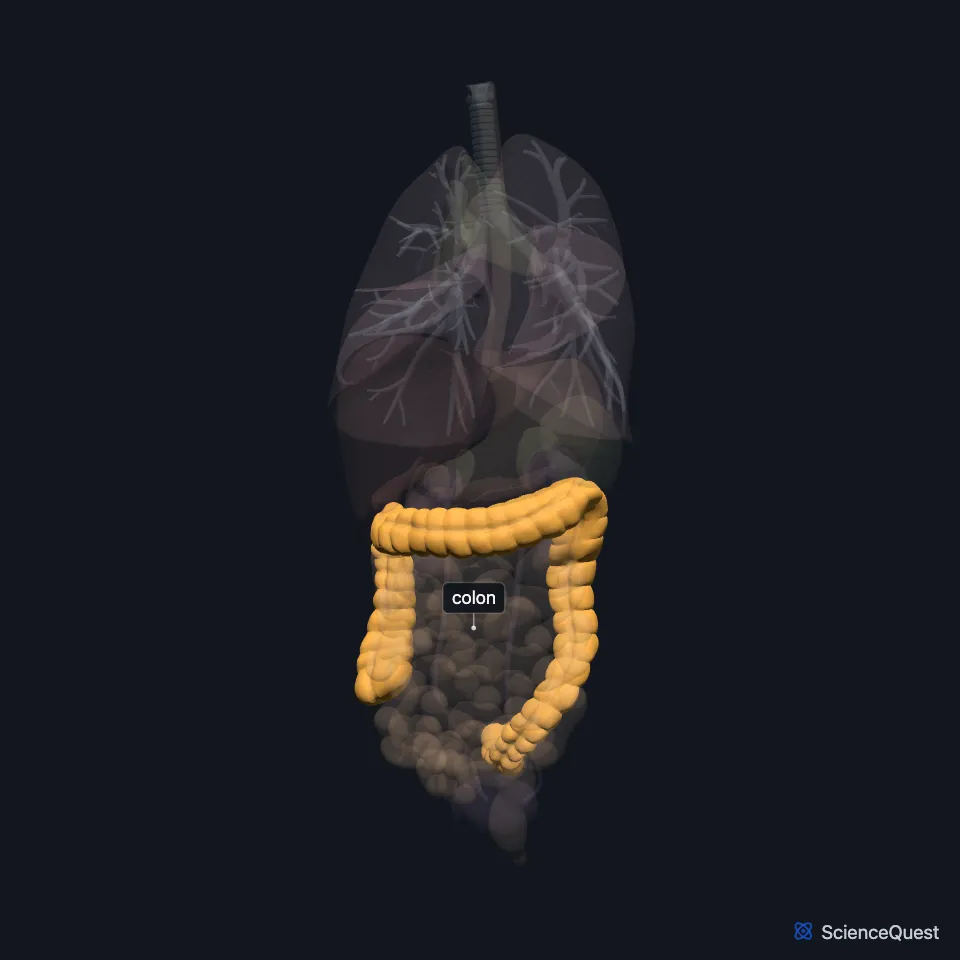
Where is the colon?
The ascending colon runs up the right side of the abdomen to the right lobe of the liver. There it turns left at the right colic (hepatic) flexure and becomes the transverse colon. (OpenStax A&P 2e, 23.5; StatPearls, Large Intestine)
The transverse colon crosses to the left and turns down below the spleen at the left colic (splenic) flexure, which the phrenicocolic ligament ties to the diaphragm. The descending colon then runs down the left side to the S-shaped sigmoid colon, which becomes the rectum at the level of S3. (OpenStax A&P 2e, 23.5; StatPearls, Large Intestine; Gray’s Anatomy (1918), The Peritoneum)
The ascending and descending colon lie behind the peritoneum. The transverse colon hangs on the transverse mesocolon, and the sigmoid colon on the sigmoid mesocolon. (OpenStax A&P 2e, 23.5; StatPearls, Large Intestine and Sigmoid Colon)
Together with the rest of the large intestine, it frames the small intestine, the jejunum and ileum. (OpenStax A&P 2e, 23.5; StatPearls, Physiology, Small Bowel)
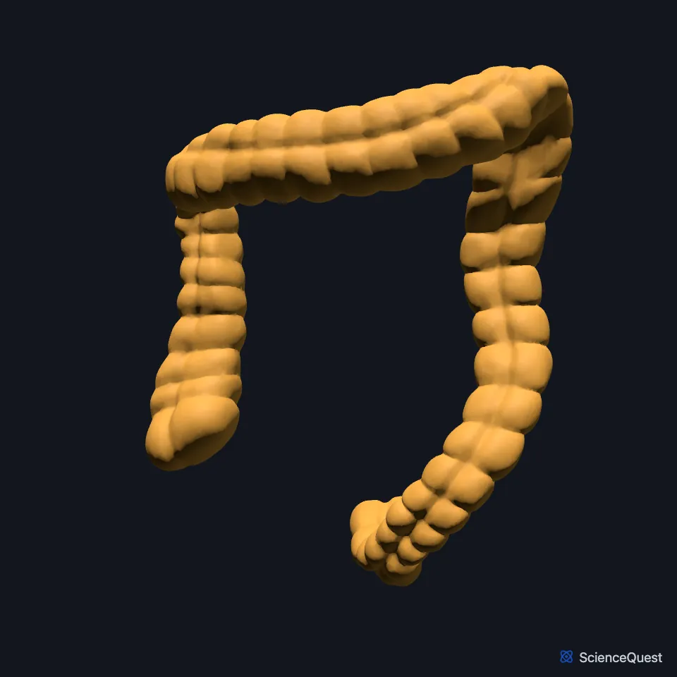
Why does the colon look puckered?
Its longitudinal muscle runs in three bands, the teniae coli. Its wall forms pouches called haustra, and small fat-filled pouches of peritoneum, the omental (epiploic) appendices, hang from it. (OpenStax A&P 2e, 23.5; StatPearls, Large Intestine)
Its lining is smooth and has no villi, unlike the lining of the small intestine. (OpenStax A&P 2e, 23.5; Gray’s Anatomy (1918), The Large Intestine)
The transverse colon is its longest and most mobile part. The large intestine as a whole is about half as long as the small intestine but more than twice as wide. (StatPearls, Large Intestine; OpenStax A&P 2e, 23.5)
What does the colon do?
By the time residue reaches the colon, the small intestine has absorbed most nutrients and up to 90% of the water. The ascending colon absorbs the remaining water and firms the residue into stool, and the descending colon stores it until it passes to the rectum. (StatPearls, Physiology, Large Intestine)
Haustral contractions push residue from one pouch to the next and mix it, which helps the colon absorb water. (OpenStax A&P 2e, 23.5; StatPearls, Physiology, Large Intestine)
Mass movements, stronger waves most active in the transverse and left colon, drive the contents toward the rectum. A meal sets them off, so you often feel the urge to defecate soon after eating. (OpenStax A&P 2e, 23.5; StatPearls, Physiology, Large Intestine and Physiology, Gastrocolic Reflex)
Bacteria in the colon make vitamins, among them vitamin K and biotin, which can be absorbed across its lining. (StatPearls, Physiology, Large Intestine; OpenStax A&P 2e, 23.5)
What supplies the colon?
The ileocolic and right colic arteries supply the ascending colon, and the transverse colon gets most of its blood from the middle colic artery. All three are branches of the superior mesenteric artery; arcades between the middle and left colic arteries add to the transverse colon’s supply. The left colic and sigmoid branches of the inferior mesenteric artery supply the descending and sigmoid colon. (StatPearls, Large Intestine)
The marginal artery runs along the whole length of the colon and links the two arterial territories. (StatPearls, Large Intestine)
The ascending colon and first two thirds of the transverse colon come from the midgut and take nerves from the superior mesenteric plexus. The last third of the transverse colon onward is hindgut and takes nerves from the inferior mesenteric plexus. (StatPearls, Large Intestine; OpenStax A&P 2e, 23.5)
Its veins drain to the portal system: the inferior mesenteric vein into the splenic vein, and the superior mesenteric vein joins the splenic vein to form the hepatic portal vein. (StatPearls, Large Intestine)
| Feature | Detail and source |
|---|---|
| Parts | Ascending, transverse, descending, sigmoid (StatPearls, Physiology, Large Intestine) |
| Flexures | Right colic (hepatic), below the liver; left colic (splenic), below the spleen (OpenStax A&P 2e, 23.5; StatPearls, Large Intestine; Gray’s Anatomy (1918), The Peritoneum) |
| Peritoneum | Ascending and descending retroperitoneal; transverse and sigmoid on a mesocolon (OpenStax A&P 2e, 23.5; StatPearls, Large Intestine and Sigmoid Colon) |
| Muscle bands | Three teniae coli (OpenStax A&P 2e, 23.5; StatPearls, Large Intestine) |
| Arteries | Superior mesenteric (ileocolic, right colic, middle colic); inferior mesenteric (left colic, sigmoid) (StatPearls, Large Intestine) |
Touches in this model
Measured on the BodyParts3D meshes: surfaces within 2 mm of the colon, nearest first, 12 of 25.
- Duodenum in contact
- Free taenia in contact
- Ileum in contact
- Jejunum in contact
- Left kidney in contact
- Liver in contact
- Mesocolic taenia in contact
- Omental taenia in contact
- Rectum in contact
- Right kidney in contact
- Spleen in contact
- Stomach in contact
Sources
- OpenStax Anatomy and Physiology 2e, section 23.5, The Small and Large Intestines. CC BY-NC-SA 4.0. Read 2026-10-25.
- StatPearls, Physiology, Large Intestine. Laura L. Azzouz; Sandeep Sharma. Last update July 31, 2023. CC BY-NC-ND 4.0. Read 2026-10-25.
- StatPearls, Anatomy, Abdomen and Pelvis: Large Intestine. Adekunle E. Omole; Pujyitha Mandiga; Preet Kahai; Stany Lobo. Last update April 6, 2025. CC BY-NC-ND 4.0. Read 2026-10-25.
- Gray’s Anatomy of the Human Body, 20th US edition (1918), Splanchnology, The Peritoneum, page 1158. Henry Gray; edited by Warren H. Lewis. Public domain. Read 2026-10-25.
- StatPearls, Anatomy, Abdomen and Pelvis, Sigmoid Colon. Joshua M. Harkins; Hussain Sajjad. Last update July 24, 2023. CC BY-NC-ND 4.0. Read 2026-10-25.
- StatPearls, Physiology, Small Bowel. Elizabeth M. Fish; Karlie R. Shumway; Bracken Burns. Last update January 31, 2024. CC BY-NC-ND 4.0. Read 2026-10-25.
- Gray’s Anatomy of the Human Body, 20th US edition (1918), Splanchnology, The Large Intestine, page 1186. Henry Gray; edited by Warren H. Lewis. Public domain. Read 2026-10-25.
- StatPearls, Physiology, Gastrocolic Reflex. Jordan C. Malone; Aravind Thavamani. Last update May 1, 2023. CC BY-NC-ND 4.0. Read 2026-10-25.
The values are facts and free to use; this page’s selection and presentation are © 2026 ScienceQuest.