Kidney
The kidneys are a pair of bean-shaped organs behind the peritoneum, one on each side of the spine between the T12 and L3 vertebrae. Each filters the blood and passes the urine it makes to a ureter. (StatPearls, Abdomen and Pelvis: Kidneys)
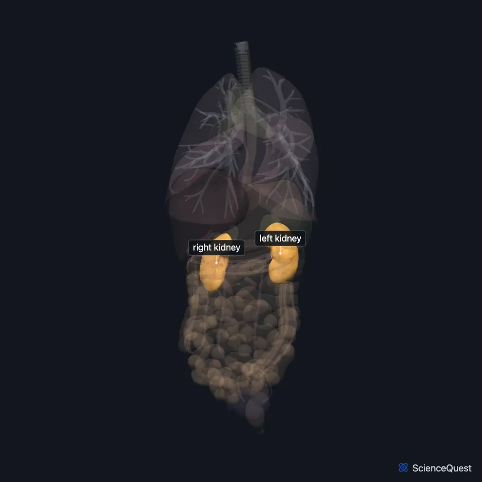
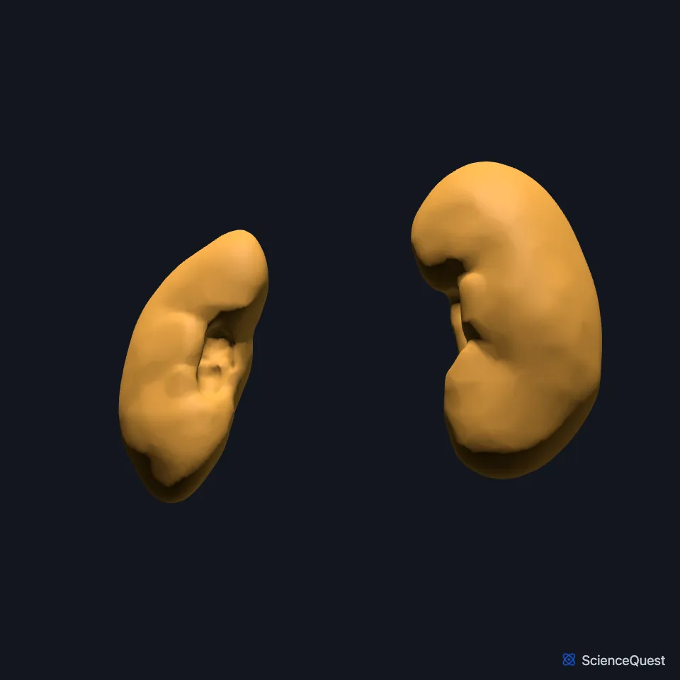
Where are the kidneys?
They lie behind the peritoneum, against the back wall of the abdomen, bedded in fat on the diaphragm and the muscles of that wall. The lower ribs shield their upper parts: the right kidney rests on the 12th rib, and the left usually on the 11th and 12th. (OpenStax A&P 2e, 25.3; StatPearls, Abdomen and Pelvis: Kidneys; Gray 1918, 3b. 1. The Kidneys)
The right kidney sits a little lower than the left, pushed down by the liver. Each kidney lies slightly oblique, parallel to the psoas major on its side, and its lower pole sits further out and further forward than its upper pole. (OpenStax A&P 2e, 25.3; StatPearls, Abdomen and Pelvis: Kidneys)
What do the kidneys do?
They excrete nitrogen wastes such as urea and ammonia, regulate sodium, potassium and calcium, and hold the acid-base balance by excreting hydrogen ions and reclaiming bicarbonate. They also help set blood pressure and blood volume through the renin-angiotensin-aldosterone system. (StatPearls, Abdomen and Pelvis: Kidneys)
When their blood flow falls they release the enzyme renin, which starts the renin-angiotensin-aldosterone system. They also make two hormones. Parathyroid hormone prompts them to make calcitriol, the active form of vitamin D, and a low oxygen level prompts them to make erythropoietin, which drives red blood cell production. (OpenStax A&P 2e, 17.10; StatPearls, Physiology, Renal, Physiology, Parathyroid Hormone, Abdomen and Pelvis: Kidneys and Erythropoietin Stimulating Agents)
Together the kidneys filter about 120 to 125 mL of plasma a minute, roughly 180 L a day. They reabsorb all but 1 to 2 L of it, and that remainder leaves as urine. (OpenStax A&P 2e, 25.5; StatPearls, Physiology, Glomerular Filtration Rate and Physiology, Renal)
What is inside a kidney?
A fibrous capsule covers each kidney, then a cushion of perinephric fat, the renal fat pad, then the tough renal fascia. The fascia and the organs pressed against the kidney hold it in place on the posterior abdominal wall. (OpenStax A&P 2e, 25.3; StatPearls, Abdomen and Pelvis: Kidneys; Gray 1918, 3b. 1. The Kidneys)
A frontal cut shows an outer cortex and an inner medulla. The medulla is made of renal pyramids separated by renal columns that run down from the cortex, and a pyramid with the cortex over it forms a renal lobe. (OpenStax A&P 2e, 25.3; StatPearls, Abdomen and Pelvis: Kidneys)
Urine leaves each pyramid at its tip, the renal papilla, into a minor calyx. Minor calyces join into major calyces, and these join into the funnel-shaped renal pelvis, where the ureter begins. (StatPearls, Abdomen and Pelvis: Kidneys)
The working unit is the nephron. It starts at the glomerulus, a tuft of capillaries enclosed by the glomerular (Bowman) capsule, and continues as a tubule through the loop of Henle to a collecting duct. (StatPearls, Abdomen and Pelvis: Kidneys)
What lies next to the kidneys?
Each kidney sits directly below its adrenal gland. Its upper pole lies in front of the diaphragm and is often crossed by the 12th rib, and its lower pole lies in front of the psoas and the outer edge of the quadratus lumborum. (StatPearls, Abdomen and Pelvis: Kidneys)
The liver lies in front of the right kidney, separated from it by the hepatorenal recess, and the second part of the duodenum lies along its inner side. The left kidney lies behind the descending colon, the tail of the pancreas, part of the stomach and the spleen, and the colon lies in front of both kidneys. (StatPearls, Abdomen and Pelvis: Kidneys)
Vessels, nerves and the ureter pass through the hilum, a deep slit in the inner border. The renal artery branches off the aorta, and the renal vein opens into the inferior vena cava. (OpenStax A&P 2e, 25.3; Gray 1918, 3b. 1. The Kidneys; StatPearls, Abdomen and Pelvis: Kidneys)
Its nerves come from the renal plexus, which carries sympathetic and parasympathetic fibers. Pain signals from the kidney reach spinal segments T11 to L2 and are often felt in the matching dermatomes, so pain in the flank can come from the kidney. (StatPearls, Abdomen and Pelvis: Kidneys)
How often are the kidneys joined?
In a horseshoe kidney the two lower poles are fused, usually in front of the aorta. It occurs in about 1 in 400 people, and these kidneys often have several accessory renal arteries. (StatPearls, Abdomen and Pelvis: Kidneys)
Where the sources differ
OpenStax gives each kidney as 11 to 14 cm long, 6 cm wide and 4 cm thick, weighing 125 to 175 g in males and 115 to 155 g in females. StatPearls gives 10 to 12 cm by 5 to 7 cm by 3 to 5 cm, with average weights of 160 to 162 g in men and 135 to 136 g in women. (OpenStax A&P 2e, 25.3; StatPearls, Abdomen and Pelvis: Kidneys)
OpenStax section 25.3 says the kidneys receive about 25% of the cardiac output at rest. OpenStax section 25.5 and StatPearls say about 20%, which StatPearls puts at 1 to 1.1 L a minute. (OpenStax A&P 2e, 25.3 and 25.5; StatPearls, Abdomen and Pelvis: Kidneys)
OpenStax lists the hilum from front to back as artery, vein, renal pelvis. The StatPearls kidney article puts the renal vein in front of the artery, with the pelvis behind both, and the StatPearls ureter article also places the start of the ureter behind the renal vein and artery. (OpenStax A&P 2e, 25.3; StatPearls, Abdomen and Pelvis: Kidneys and Abdomen and Pelvis Ureter)
OpenStax counts about 1.3 million nephrons in each kidney; StatPearls gives 1 to 1.5 million, occasionally more than 2.5 million. (OpenStax A&P 2e, 25.3; StatPearls, Abdomen and Pelvis: Kidneys)
| Feature | Detail and source |
|---|---|
| Vertebral level | T12 to L3 (StatPearls, Abdomen and Pelvis: Kidneys) |
| Artery | Renal artery, from the aorta, entering the hilum at L2 (StatPearls, Abdomen and Pelvis: Kidneys) |
| Drains urine to | Ureter, through the renal pelvis (StatPearls, Abdomen and Pelvis: Kidneys) |
| Nerves | Renal plexus, sympathetic and parasympathetic (StatPearls, Abdomen and Pelvis: Kidneys) |
Touches in this model
Measured on the BodyParts3D meshes: surfaces within 2 mm of the kidney, nearest first, 12 of 25.
- Colon in contact
- Left adrenal gland in contact
- Left ureter in contact
- Liver in contact
- Mesocolic taenia in contact
- Omental taenia in contact
- Right adrenal gland in contact
- Right ureter in contact
- Spleen in contact
- Diaphragm in contact
- Internal intercostal muscle in contact
- Left psoas major in contact
Sources
- StatPearls, Anatomy, Abdomen and Pelvis: Kidneys. Felix A. Garza; Stephen W. Leslie. Last update September 15, 2025. CC BY-NC-ND 4.0. Read 2026-10-25.
- OpenStax Anatomy and Physiology 2e, section 25.3, Gross Anatomy of the Kidney. CC BY-NC-SA 4.0. Read 2026-10-25.
- Gray’s Anatomy of the Human Body, 20th US edition (1918), XI. Splanchnology, 3b. The Urinary Organs, 1. The Kidneys (1918 terms: adipose capsule for perinephric fat, fibrous tunic for renal capsule). Henry Gray. Public domain. Read 2026-10-25.
- OpenStax Anatomy and Physiology 2e, section 17.10, Organs with Secondary Endocrine Functions. CC BY-NC-SA 4.0. Read 2026-10-25.
- StatPearls, Physiology, Renal. Ifeanyichukwu Ogobuiro; Faiz Tuma. Last update July 24, 2023. CC BY-NC-ND 4.0. Read 2026-10-25.
- StatPearls, Physiology, Parathyroid Hormone. Maqsood Khan; Alvin Jose; Sandeep Sharma. Last update October 29, 2022. CC BY-NC-ND 4.0. Read 2026-10-25.
- StatPearls, Erythropoietin Stimulating Agents. Benjamin Schoener; Judith Borger. Last update July 14, 2024. CC BY-NC-ND 4.0. Read 2026-10-25.
- OpenStax Anatomy and Physiology 2e, section 25.5, Physiology of Urine Formation. CC BY-NC-SA 4.0. Read 2026-10-25.
- StatPearls, Physiology, Glomerular Filtration Rate. Daniel P. Kaufman; Hajira Basit; Stephen J. Knohl. Last update July 17, 2023. CC BY-NC-ND 4.0. Read 2026-10-25.
- StatPearls, Anatomy, Abdomen and Pelvis Ureter. Hernan A. Lescay; Jay Jiang; Stephen W. Leslie; Faiz Tuma. Last update May 5, 2024. CC BY-NC-ND 4.0. Read 2026-10-25.
The values are facts and free to use; this page’s selection and presentation are © 2026 ScienceQuest.