Renal artery
The renal arteries are the only blood supply of the kidneys. Each leaves the side of the abdominal aorta, usually at the level of the disc between L1 and L2. (StatPearls, Abdomen and Pelvis, Renal Artery)
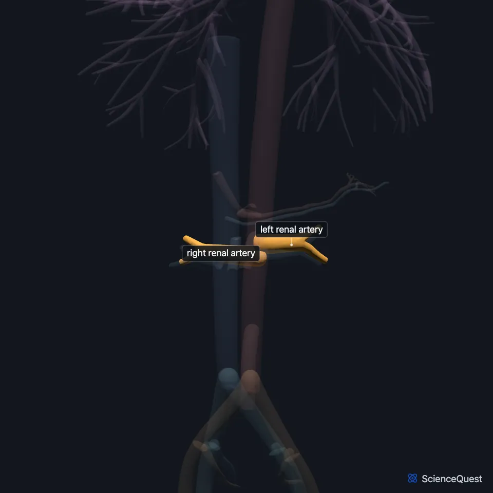
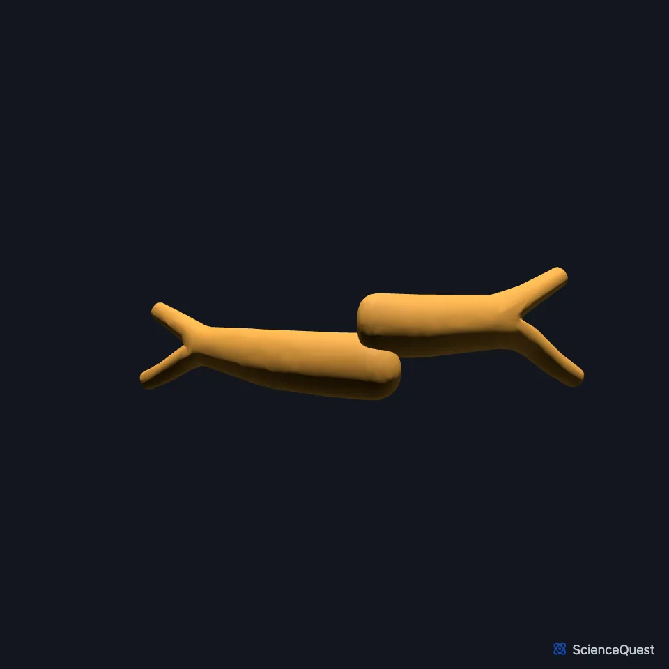
Where are the renal arteries?
Each is about 4 to 6 cm long and 5 to 6 mm wide. They run outward and backward to the hilum of the kidney. (StatPearls, Abdomen and Pelvis, Renal Artery)
The right renal artery is the longer of the two. OpenStax puts this down to the aorta lying left of the vertebral column, so the right artery has further to travel. It passes behind the inferior vena cava to reach the right kidney. (OpenStax A&P 2e, 20.5; StatPearls, Abdomen and Pelvis, Renal Artery)
What does the renal artery supply?
At rest the kidneys take a large share of the cardiac output: about a quarter in OpenStax, and about 20%, roughly 1 liter per minute, in StatPearls. (OpenStax A&P 2e, 25.3; StatPearls, Physiology, Renal Blood Flow and Filtration)
The renal artery also supplies the adrenal gland and the ureter on its own side. (StatPearls, Abdomen and Pelvis, Renal Artery)
How does the renal artery branch?
Before the hilum it splits into an anterior and a posterior division, which carry about 75% and 25% of its blood. (StatPearls, Abdomen and Pelvis, Renal Artery)
Inside the kidney it branches into segmental, interlobar, arcuate and cortical radiate (interlobular) arteries, then into afferent arterioles. Each adult kidney holds about 1 to 1.5 million nephrons; OpenStax gives 1.3 million. (OpenStax A&P 2e, 25.3; StatPearls, Abdomen and Pelvis, Renal Artery and Abdomen and Pelvis: Kidneys)
What lies next to the renal artery?
Each kidney connects directly to the great vessels: the renal artery is a branch of the aorta itself, and the renal vein ends in the inferior vena cava. The artery enters the kidney, and the vein and renal pelvis leave it, at the renal hilum. (OpenStax A&P 2e, 25.3; StatPearls, Abdomen and Pelvis, Renal Artery, Abdomen and Pelvis, Renal Veins and Abdomen and Pelvis: Kidneys)
Each artery runs behind its renal vein and enters the hilum in front of the renal pelvis. Behind the first part of the right artery lie the right crus of the diaphragm and the psoas major. (StatPearls, Abdomen and Pelvis, Renal Artery)
How common are extra renal arteries?
At least 30% of people have accessory renal arteries, and about 10% have a renal artery that branches early. (StatPearls, Abdomen and Pelvis, Renal Artery)
Where the sources differ
OpenStax places the renal arteries about 2.5 cm below the superior mesenteric artery; StatPearls has them arise immediately below it. (OpenStax A&P 2e, 20.5; StatPearls, Abdomen and Pelvis, Renal Artery)
OpenStax lists the order at the hilum as artery, vein, renal pelvis from front to back, while StatPearls puts the artery behind the renal vein. (OpenStax A&P 2e, 25.3; StatPearls, Abdomen and Pelvis, Renal Artery)
OpenStax places the aorta to the left of the vertebral column. StatPearls (Abdominal Aorta) places the abdominal aorta in front of the lumbar vertebrae, slightly to the left, with the inferior vena cava to its right. (OpenStax A&P 2e, 20.5; StatPearls, Abdomen and Pelvis: Abdominal Aorta)
| Feature | Detail and source |
|---|---|
| Arises from | Side of the abdominal aorta, usually at the L1/L2 disc (StatPearls, Abdomen and Pelvis, Renal Artery) |
| Length and width | 4 to 6 cm long, 5 to 6 mm wide (StatPearls, Abdomen and Pelvis, Renal Artery) |
| Enters | Renal hilum, behind the renal vein and in front of the renal pelvis (StatPearls, Abdomen and Pelvis, Renal Artery) |
Touches in this model
Measured on the BodyParts3D meshes: surfaces within 2 mm of the renal artery, nearest first, 12 of 15.
- Descending aorta in contact
- Inferior vena cava in contact
- Left renal vein in contact
- Right renal vein in contact
- Superior mesenteric artery in contact
- Diaphragm in contact
- First lumbar vertebra in contact
- Intervertebral disk of first lumbar vertebra in contact
- Left kidney in contact
- Left ureter in contact
- Right kidney in contact
- Right psoas major in contact
Sources
- StatPearls, Anatomy, Abdomen and Pelvis, Renal Artery. Stephen W. Leslie; Hussain Sajjad. Last update August 8, 2023. CC BY-NC-ND 4.0. Read 2026-10-25.
- OpenStax Anatomy and Physiology 2e, section 20.5, Circulatory Pathways. CC BY-NC-SA 4.0. Read 2026-10-25.
- OpenStax Anatomy and Physiology 2e, section 25.3, Gross Anatomy of the Kidney. CC BY-NC-SA 4.0. Read 2026-10-25.
- StatPearls, Physiology, Renal Blood Flow and Filtration. Rajeev Dalal; Zachary S. Bruss; Jasjit S. Sehdev. Last update July 24, 2023. CC BY-NC-ND 4.0. Read 2026-10-25.
- StatPearls, Anatomy, Abdomen and Pelvis: Kidneys. Felix A. Garza; Stephen W. Leslie. Last update September 15, 2025. CC BY-NC-ND 4.0. Read 2026-10-25.
- StatPearls, Anatomy, Abdomen and Pelvis, Renal Veins. Cole S. Bowdino; Justin Owens; Palma M. Shaw. Last update January 2, 2023. CC BY-NC-ND 4.0. Read 2026-10-25.
- StatPearls, Anatomy, Abdomen and Pelvis: Abdominal Aorta. Clement T. Tran; Cheng Ying Wu; Stephen J. Bordes; Forshing Lui. Last update July 25, 2023. CC BY-NC-ND 4.0. Read 2026-10-25.
The values are facts and free to use; this page’s selection and presentation are © 2026 ScienceQuest.