Ureter
The ureters are two thin muscular tubes, 3 to 4 mm across, that carry urine from the renal pelvis of each kidney to the urinary bladder. (StatPearls, Abdomen and Pelvis Ureter)
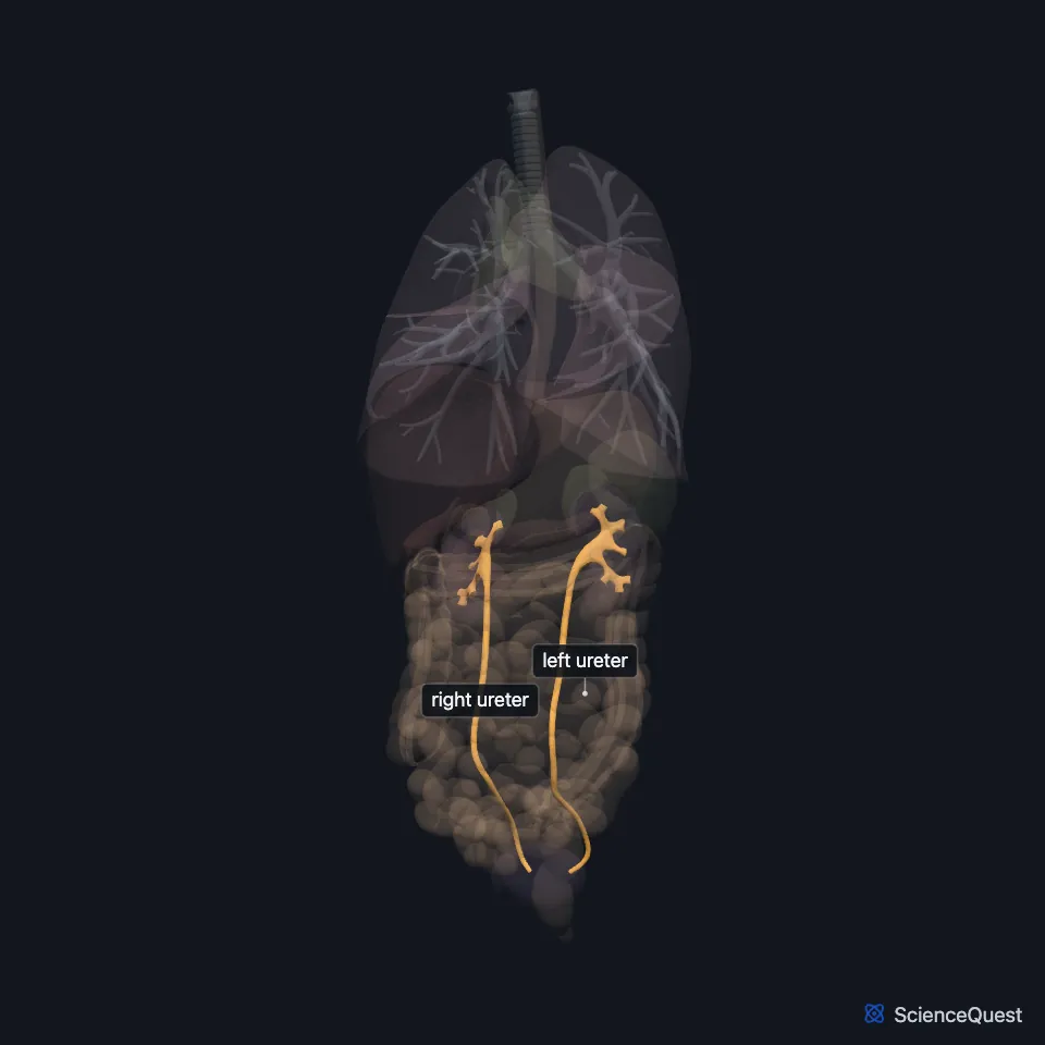
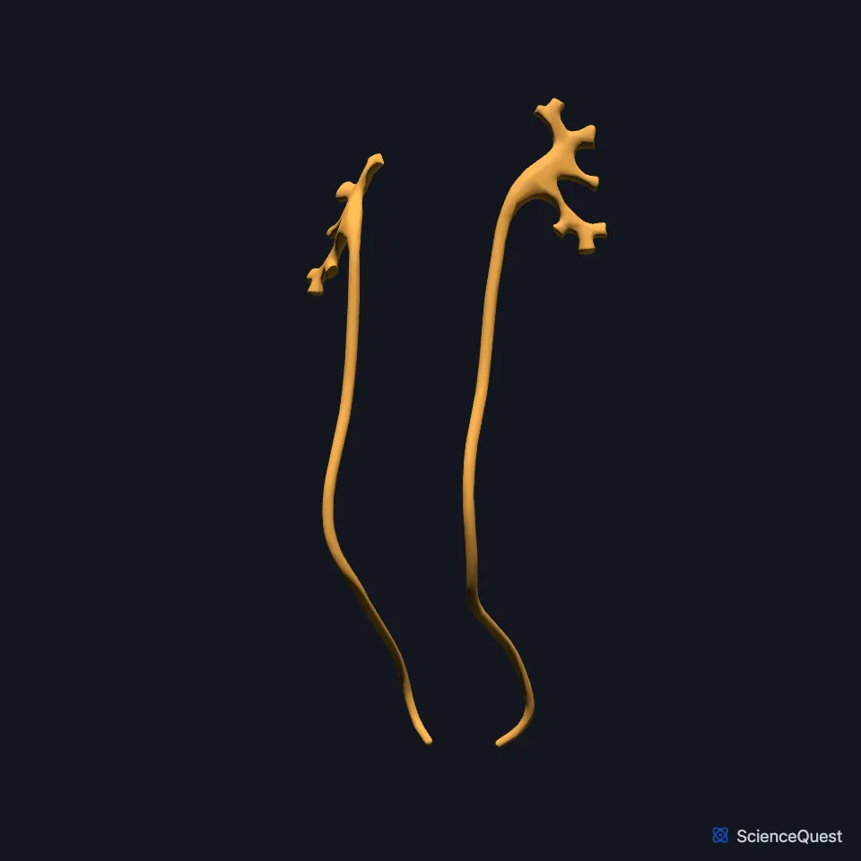
Where do the ureters run?
Each ureter starts at the ureteropelvic junction, roughly at the level of L2, and runs down behind the peritoneum in front of the psoas major. It crosses into the pelvis over the bifurcation of the common iliac artery, then follows the side wall of the pelvis to the bladder. (StatPearls, Abdomen and Pelvis Ureter)
In the pelvis the ureters sweep outward along the pelvic walls, then turn inward and pierce the bladder wall at a slant. They enter the bladder from behind and to the side, through the trigone. (OpenStax A&P 2e, 25.2; StatPearls, Abdomen and Pelvis Ureter)
How do the ureters move urine?
Waves of peristalsis in its smooth muscle, which runs in longitudinal and circular layers, push urine from the kidney down to the bladder. (OpenStax A&P 2e, 25.2; StatPearls, Abdomen and Pelvis Ureter)
The pacemaker cells that start each wave lie in the minor calyces. The wave fades at the ureterovesical junction, where the circular muscle layer ends. (StatPearls, Abdomen and Pelvis Ureter)
The slanted path through the bladder wall works as a one-way valve. It lets urine into the bladder and stops it flowing back up the ureter when the bladder contracts. (OpenStax A&P 2e, 25.2; StatPearls, Abdomen and Pelvis Ureter)
Where does the ureter narrow?
It narrows where it leaves the renal pelvis and where it enters the bladder, and stones can lodge at both points. The sharp bend at the pelvic brim is not a true narrowing, but long stones can struggle to pass it. (StatPearls, Abdomen and Pelvis Ureter)
The last part, inside the bladder wall, is about 2 cm long, has no peristalsis and is the narrowest part of the ureter. (StatPearls, Abdomen and Pelvis Ureter)
Transitional epithelium over a thick lamina propria lines it and makes a watertight barrier, and it has no submucosa. Outside the muscle coat, a fibroelastic adventitia carries its vessels, lymphatics and nerves. (StatPearls, Abdomen and Pelvis Ureter)
What lies next to the ureters?
The gonadal vessels cross in front of each ureter about a third of the way to the bladder. The genitofemoral nerve lies on the psoas just behind the upper ureter. (StatPearls, Abdomen and Pelvis Ureter)
The right ureter lies near the ascending colon, cecum and appendix, and the left lies close to the descending and sigmoid colon. (StatPearls, Abdomen and Pelvis Ureter)
In men each ureter passes behind the deferent duct and in front of the seminal vesicle just before it enters the bladder. (StatPearls, Abdomen and Pelvis Ureter)
Its blood supply comes in segments. The renal artery feeds the part nearest the kidney, branches of the aorta and the common iliac and gonadal arteries feed the middle, and branches of the internal iliac artery feed the lowest part. (StatPearls, Abdomen and Pelvis Ureter)
Where the sources differ
OpenStax gives the ureters as about 30 cm long. StatPearls gives an average of 26 cm, with a range of 22 to 30 cm. (OpenStax A&P 2e, 25.2; StatPearls, Abdomen and Pelvis Ureter)
| Feature | Detail and source |
|---|---|
| Diameter | 3 to 4 mm (StatPearls, Abdomen and Pelvis Ureter) |
| Runs from | Renal pelvis, at the ureteropelvic junction, about L2 (StatPearls, Abdomen and Pelvis Ureter) |
| Ends in | Urinary bladder, through the trigone (StatPearls, Abdomen and Pelvis Ureter) |
| Lining | Transitional epithelium (OpenStax A&P 2e, 25.2; StatPearls, Abdomen and Pelvis Ureter) |
Touches in this model
Measured on the BodyParts3D meshes: surfaces within 2 mm of the ureter, nearest first, 12 of 20.
- Left kidney in contact
- Right kidney in contact
- Urinary bladder in contact
- Left common iliac artery in contact
- Left deferent duct in contact
- Left external iliac vein in contact
- Left psoas major in contact
- Left renal artery in contact
- Left renal vein in contact
- Right common iliac artery in contact
- Right deferent duct in contact
- Right external iliac vein in contact
Sources
- StatPearls, Anatomy, Abdomen and Pelvis Ureter. Hernan A. Lescay; Jay Jiang; Stephen W. Leslie; Faiz Tuma. Last update May 5, 2024. CC BY-NC-ND 4.0. Read 2026-10-25.
- OpenStax Anatomy and Physiology 2e, section 25.2, Gross Anatomy of Urine Transport. CC BY-NC-SA 4.0. Read 2026-10-25.
The values are facts and free to use; this page’s selection and presentation are © 2026 ScienceQuest.