Seminal vesicle
The seminal vesicles are a pair of glands in the male pelvis. Their fluid, rich in fructose, makes up most of the volume of semen. (StatPearls, Abdomen and Pelvis, Seminal Vesicle; OpenStax A&P 2e, 27.1)
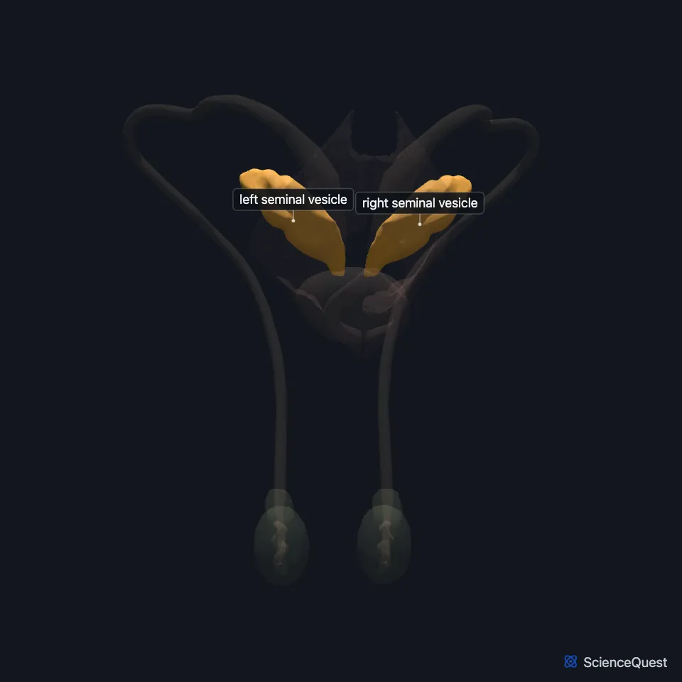
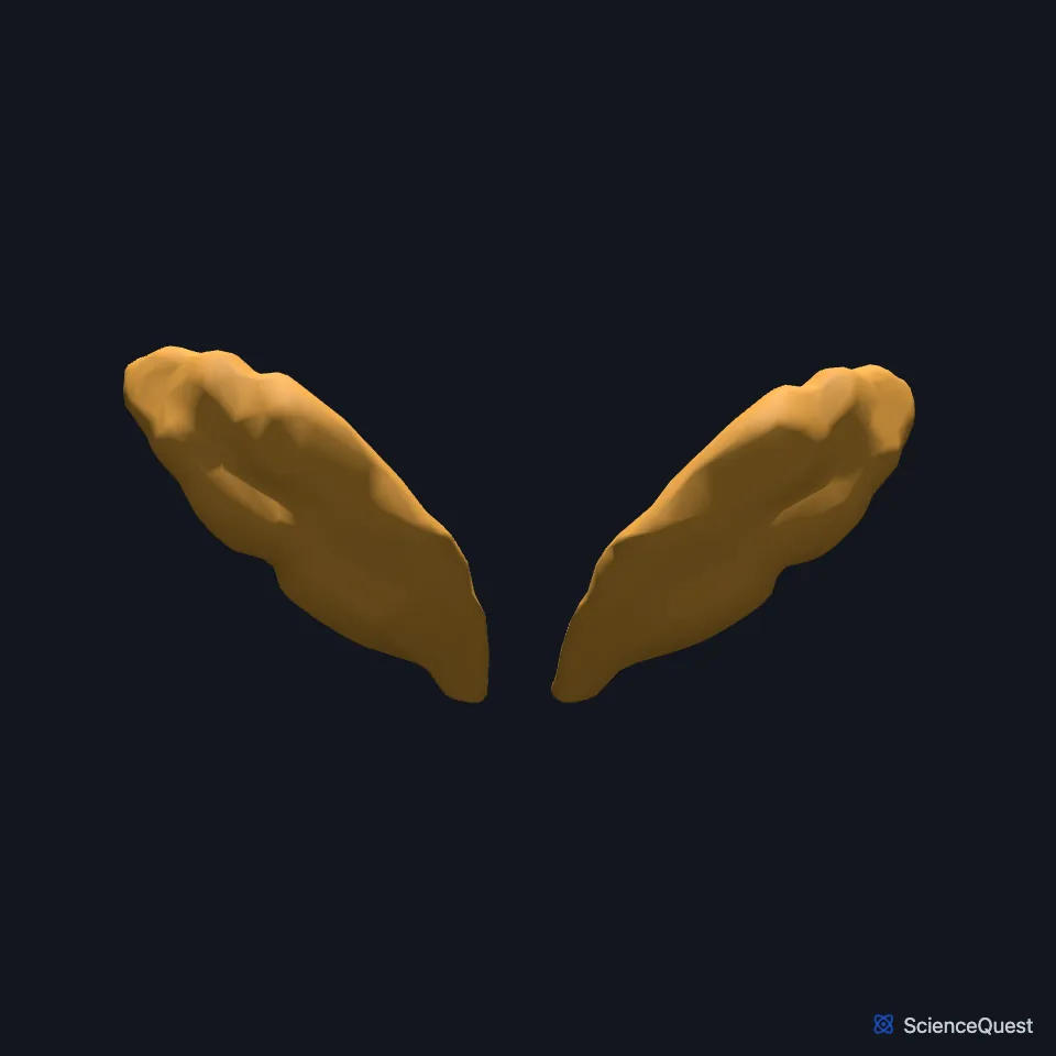
Open it in the Male Reproductive Organs Explorer See it alone in 3D
Where are the seminal vesicles?
Each lies to the outer side of the ampulla of a deferent duct. Like the prostate, they are separated from the rectum by the rectovesical septum (Denonvilliers fascia). (StatPearls, Abdomen and Pelvis, Seminal Vesicle)
What is a seminal vesicle made of?
Each is a single coiled tube, closed at one end, with several irregular pouches. It is usually 3 to 5 cm long and 1 cm across, and about 10 cm long when uncoiled. (StatPearls, Abdomen and Pelvis, Seminal Vesicle)
The wall has three layers: a mucosa of pseudostratified columnar epithelium, a smooth muscle layer with inner circular and outer longitudinal fibers, and an outer adventitia of loose areolar tissue. (StatPearls, Abdomen and Pelvis, Seminal Vesicle)
They grow out of the lower mesonephric ducts in the tenth week of fetal life, in response to testosterone. The prostate and bulbourethral glands, by contrast, come from the urogenital sinus under dihydrotestosterone. (StatPearls, Abdomen and Pelvis, Seminal Vesicle)
What do the seminal vesicles do?
Their fluid is slightly alkaline, which raises the pH of semen against the acid of the vagina. It also carries fructose, which sperm use to make ATP for swimming. (StatPearls, Abdomen and Pelvis, Seminal Vesicle; OpenStax A&P 2e, 27.1)
Their protein semenogelin forms a gel around ejaculated sperm. Prostate-specific antigen later breaks the gel down and frees them. (StatPearls, Abdomen and Pelvis, Seminal Vesicle)
In the emission phase of ejaculation, sympathetic fibers in the hypogastric nerves make the seminal vesicles and prostate contract. That pushes sperm and seminal fluid into the posterior urethra. (StatPearls, Abdomen and Pelvis, Seminal Vesicle)
What lies next to the seminal vesicles?
Each seminal vesicle joins the ampulla of its deferent duct to form an ejaculatory duct. The two ejaculatory ducts open into the prostatic urethra at the seminal colliculus (verumontanum), one on each side of the prostatic utricle. (StatPearls, Abdomen and Pelvis, Seminal Vesicle)
In men each ureter passes in front of the seminal vesicle just before it enters the bladder. (StatPearls, Abdomen and Pelvis Ureter)
Their arteries come from the middle and inferior vesical and middle rectal vessels. Veins drain through the vesical venous plexus to the internal iliac veins, and nerves come from the hypogastric nerves and the inferior hypogastric plexus. (StatPearls, Abdomen and Pelvis, Seminal Vesicle)
Where the sources differ
OpenStax says the seminal vesicles contribute about 60% of semen volume. StatPearls says about 70%. (OpenStax A&P 2e, 27.1; StatPearls, Abdomen and Pelvis, Seminal Vesicle)
OpenStax says prostatic fluid first thickens semen and then thins it. StatPearls says the gel forms from semenogelin made by the seminal vesicles, and prostate-specific antigen then liquefies it. (OpenStax A&P 2e, 27.1; StatPearls, Abdomen and Pelvis, Seminal Vesicle)
| Feature | Detail and source |
|---|---|
| Size | 3 to 5 cm long, 1 cm across; about 10 cm uncoiled (StatPearls, Abdomen and Pelvis, Seminal Vesicle) |
| Duct joins | Ampulla of the deferent duct, forming the ejaculatory duct (StatPearls, Abdomen and Pelvis, Seminal Vesicle) |
| Fluid | Slightly alkaline, with fructose and semenogelin (StatPearls, Abdomen and Pelvis, Seminal Vesicle) |
| Nerves | Hypogastric nerves and inferior hypogastric plexus (StatPearls, Abdomen and Pelvis, Seminal Vesicle) |
Touches in this model
Measured on the BodyParts3D meshes: surfaces within 2 mm of the seminal vesicle, nearest first.
- Left deferent duct in contact
- Left pubococcygeus in contact
- Prostate in contact
- Right deferent duct in contact
- Right pubococcygeus in contact
- Left iliococcygeus in contact
- Rectum in contact
- Right iliococcygeus in contact
- Skin in contact
- Urinary bladder in contact
- Right ureter 0.2 mm away
Sources
- StatPearls, Anatomy, Abdomen and Pelvis, Seminal Vesicle. Alastair C. McKay; Nkiruka Odeluga; Jay Jiang; Sandeep Sharma. Last update July 24, 2023. CC BY-NC-ND 4.0. Read 2026-10-25.
- OpenStax Anatomy and Physiology 2e, section 27.1, Anatomy and Physiology of the Testicular Reproductive System. CC BY-NC-SA 4.0. Read 2026-10-25.
- StatPearls, Anatomy, Abdomen and Pelvis Ureter. Hernan A. Lescay; Jay Jiang; Stephen W. Leslie; Faiz Tuma. Last update May 5, 2024. CC BY-NC-ND 4.0. Read 2026-10-25.
The values are facts and free to use; this page’s selection and presentation are © 2026 ScienceQuest.