Teeth Explorer
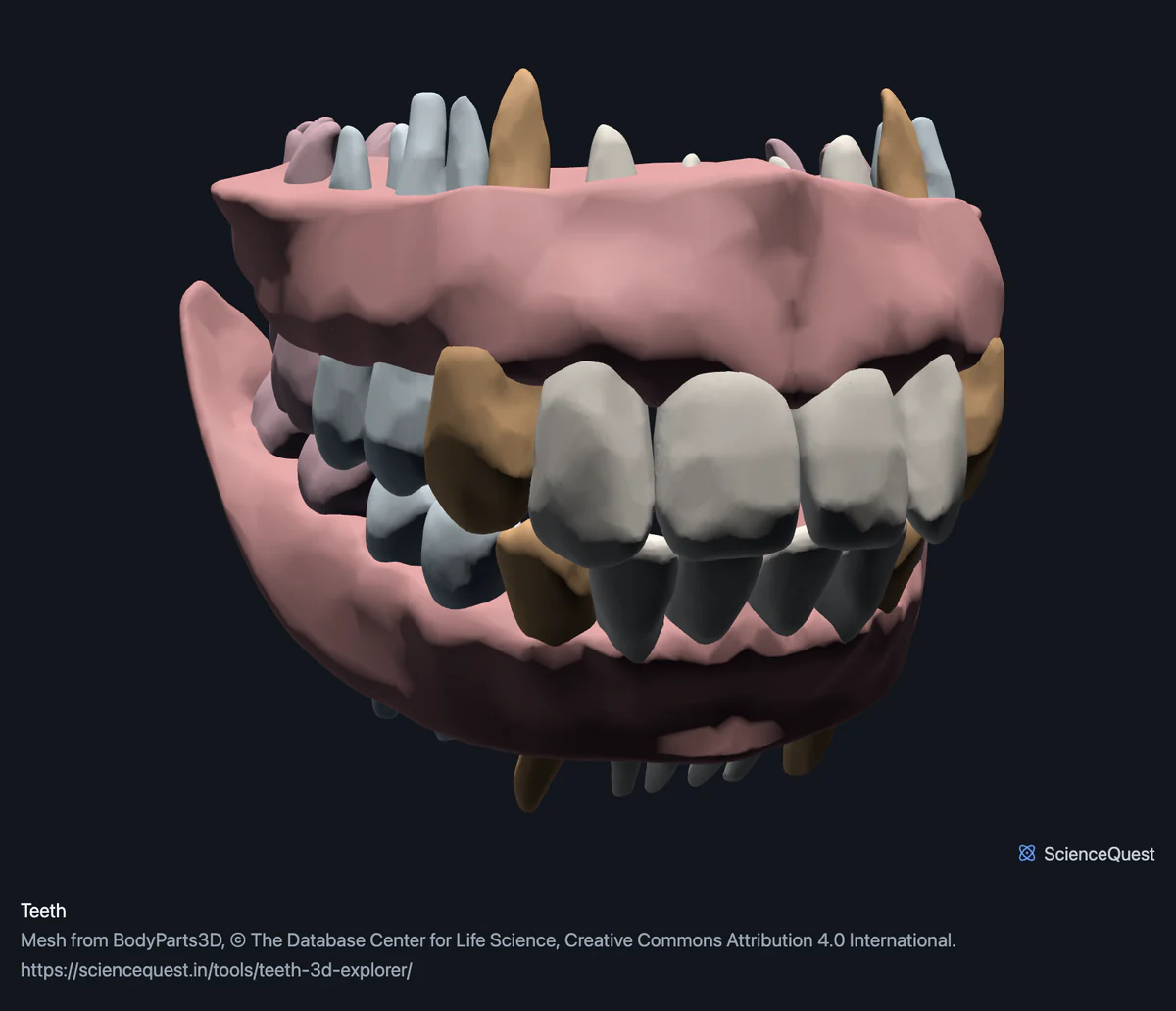
Turn a real permanent dentition in 3D: every incisor, canine, premolar and molar as its own mesh, named by position, with the gums of both jaws.
Visualiser
Teeth, 30 structures
Checking this device for a stored copy.
Start here
- Hide the Gums, then turn to a side view The roots are the surprise. The gums here are solid and enclose most of every root, but there is no jaw bone, so with them hidden every root is exposed along its whole length, and each tooth turns out to be mostly root: more than half, and on the molars there are two or three of them.
- Click Molars twice, then Below Opening a band frames it, and this one starts open, so the second click frames the molars. Below then looks up at them from underneath. The teeth are modelled with the jaws closed, so the lower molars hide most of the upper chewing surfaces, and with the gums hidden what you see are the lower molars’ roots, two to each tooth. Molars have four or five cusps, premolars two or three.
- Select one tooth, then read its dimensions Every measurement comes from the mesh, so you can compare a central incisor against a first molar in millimetres rather than by eye.
Teeth and gums. No third molars, because this person did not have them. No milk teeth, no jaw bones, no periodontal ligament, no pulp cavity and no enamel and dentine as separate layers: each tooth is one solid surface.
- 30 structures
- 218k triangles
- 1200 KB, on request
- 0.000543 mm worst error per axis
- 8 incisors
- 4 canines
- 8 premolars
- 8 molars
- 2 gums
28 teeth, within 30 meshes. A full adult dentition is 32. This body has no third molars, which is common, so 28 is a complete mouth for this specimen rather than the textbook figure. The remaining two meshes are the gums of the upper and lower jaw.
Where this comes from
Segmented from imaging of one adult male body, not drawn by an artist, so the shapes are measurements. Two consequences worth knowing: it is one person’s anatomy rather than an average, and faint stepping on some surfaces is the resolution of the original scan rather than a rendering fault.
Meshes from BodyParts3D, © The Database Center for Life Science, under Creative Commons Attribution 4.0 International. Names and identifiers are the dataset’s own: Foundational Model of Anatomy identifiers and English labels, as its parts list gives them. Converted meshes are shared under the same licence.
Changes made: Meshes were converted from binary STL to an indexed mesh with 16-bit quantised vertex positions, and surface normals were recomputed from the geometry. No vertex was moved other than by that quantisation, whose worst-case error on any one coordinate is reported on the page.
Latin terms for 0 of the 30 structures come from English Wikipedia anatomy infoboxes, under Creative Commons Attribution-ShareAlike 4.0, joined by Wikidata property P1402, Foundational Model of Anatomy identifier. Only a term a source states for that structure is used, never one constructed from a family name, which is why the rest carry no Latin rather than a guess.
All 30 structures
Every structure the model contains, group by group. Each name opens the viewer with that structure selected, so a link can point at one structure rather than at the whole region.
Incisors 8
- right upper central secondary incisor tooth tooth
- left upper central secondary incisor tooth tooth
- right upper lateral secondary incisor tooth tooth
- left upper lateral secondary incisor tooth tooth
- right lower central secondary incisor tooth tooth
- left lower central secondary incisor tooth tooth
- right lower lateral secondary incisor tooth tooth
- left lower lateral secondary incisor tooth tooth
Canines 4
Premolars 8
- right upper first secondary premolar tooth tooth
- left upper first secondary premolar tooth tooth
- right upper second secondary premolar tooth tooth
- left upper second secondary premolar tooth tooth
- right lower first secondary premolar tooth tooth
- left lower first secondary premolar tooth tooth
- right lower second secondary premolar tooth tooth
- left lower second secondary premolar tooth tooth
Molars 8
- right upper first secondary molar tooth tooth
- left upper first secondary molar tooth tooth
- right upper second secondary molar tooth tooth
- left upper second secondary molar tooth tooth
- right lower first secondary molar tooth tooth
- left lower first secondary molar tooth tooth
- right lower second secondary molar tooth tooth
- left lower second secondary molar tooth tooth
Gums 2
- gingiva of upper jaw other tissue
- gingiva of lower jaw other tissue
Citing this tool
Last updated . Add the date you accessed it as well, which a citation of a page that can change asks for. If a specific view matters, cite the page address as it stands in your browser instead: it records what is selected and shown.
Twenty eight teeth, and why that is complete
A full adult dentition is 32 teeth counting the third molars, and 28 without them. This dataset publishes first and second molars in each quadrant and no thirds, so this person either never developed wisdom teeth or had them removed before the imaging.
Both counts are normal. Roughly a quarter of people are missing at least one third molar, and many more have had them out. So 28 here is a complete mouth for one specimen rather than a dataset with four teeth missing, and the practical consequence is simple: this model cannot show you a wisdom tooth.
Each quadrant has seven
Central and lateral incisors, a canine, first and second premolars, and first and second molars. Four quadrants of seven is the 28. The names are the dataset’s own, so a tooth reads as right upper first secondary molar tooth, where secondary means permanent as opposed to the primary or milk dentition, which is not in this data.
Most of a tooth is not in the mouth
This is what the model shows that a diagram of a dental chart cannot. Each mesh is a whole tooth, root included, and the roots are longer than the crowns. An upper first molar has three roots, the lower ones have two, and the canine roots are the longest in the mouth, which is why canines are among the hardest teeth to extract.
In this model the gums are not a thin cuff at the necks of the teeth. The dataset publishes each gingiva as a solid block that encloses most of every root, and there is no alveolar bone here at all, so hide the Gums and the roots stand fully exposed in a way they never are in a living mouth. Treat the gum line as the marker of where a tooth becomes visible, and everything below it as normally buried in bone that this model does not carry.
The jaw is in a different model, on purpose
The mandible sits under the same node in the dataset and is not here, because it is a bone and it belongs with the skull and the joint it moves on. Every region here shares one coordinate frame, so opening the skull model beside this one puts these teeth in their real sockets rather than in two drawings at two scales.
One limit worth stating: each tooth is a single solid surface. There is no enamel and dentine as separate layers, no pulp cavity and no periodontal ligament, so this shows the shape of a tooth and not its internal structure.
Common questions
Why are there 28 teeth and not 32?
Because this person had 28. The dataset publishes first and second molars in each quadrant and no third molars, so either the wisdom teeth never developed or they had been removed before the imaging. A full adult dentition is 32 counting third molars and 28 without them, and both numbers are normal: roughly a quarter of people are missing at least one third molar. So this is a complete mouth for this specimen rather than a dataset with four teeth missing, and the model cannot show you a wisdom tooth.
How are the teeth named?
By quadrant, jaw and position, exactly as the dataset labels them, so a molar reads as right upper first secondary molar tooth. Secondary means permanent, as opposed to the primary or milk dentition, which is not in this data. Each quadrant has seven teeth here: central and lateral incisors, a canine, first and second premolars, and first and second molars.
Is the jaw in here?
No, and that is deliberate. The mandible sits under the same node in the dataset, but it is a bone and it ships with the skull model, where it belongs with the joint it moves on. Putting a single bone in a set of teeth would also make the page lead with a count of one bone, since the headline figure reports bone whenever a region contains any. Open the skull model alongside this one: they share a coordinate frame, so the teeth sit in their real sockets.
Are the roots modelled?
Yes, and it is the main reason to look at this in 3D rather than at a chart. Each tooth is a whole tooth, so the crown you would see in a mouth is a minority of the mesh: the roots are longer than the crowns and the molars have two or three each. Worth knowing before you read the model: there is no jaw bone in it, and the gums are solid blocks that enclose most of each root, so hide the Gums and every root stands exposed in a way it never is in a living mouth.