Knee Skeleton Explorer
Turn the four bones of the knee in 3D. See why the patella sits in front of the femur and why the fibula is not part of the joint at all.
Visualiser
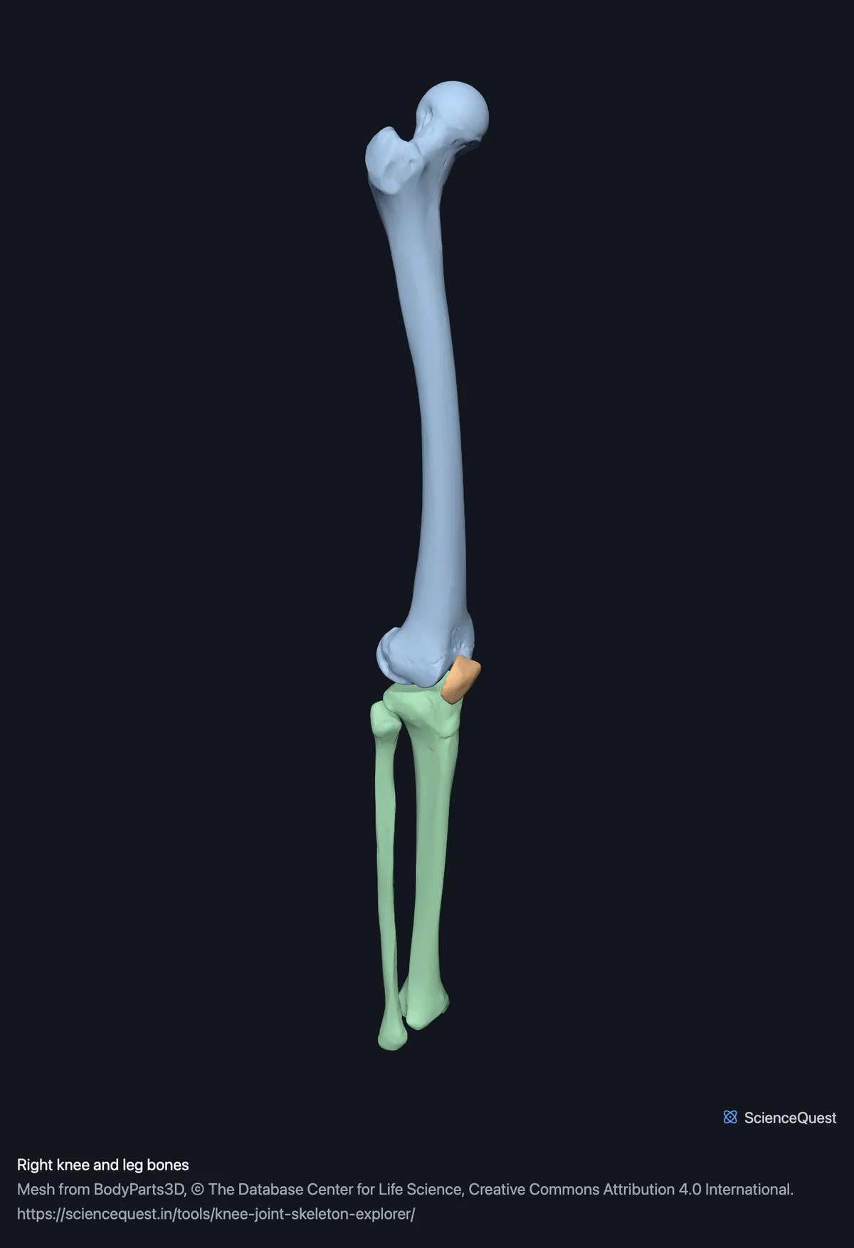
Right knee and leg bones, 4 bones
Checking this device for a stored copy.
Start here
- Turn to Lateral Shows the patella sitting in front of the femur, its centre about 20 mm forward of the line of the femoral shaft, which is the whole reason it exists.
- Isolate the fibula Its head sits below the joint line, so it is not part of the knee at all.
- Compare Front and Back, then press Recentre The medial condyle reaches further down than the lateral one. Recentre gets the whole 800 mm span back after zooming.
Bone only. The menisci, the cruciates and the collaterals are what most people come to a knee model for, and BodyParts3D does not publish them: there is no cruciate, meniscus or articular disc anywhere in its parts list, at any size. Treat this as the bony frame they attach to.
- 4 bones
- 26k triangles
- 170 KB, on request
- 0.00612 mm worst error per axis
- 1 thigh
- 1 sesamoid
- 2 lower leg
Where this comes from
Segmented from imaging of one adult male body, not drawn by an artist, so the shapes are measurements. Two consequences worth knowing: it is one person’s anatomy rather than an average, and faint stepping on some surfaces is the resolution of the original scan rather than a rendering fault.
Meshes from BodyParts3D, © The Database Center for Life Science, under Creative Commons Attribution 4.0 International. Names and identifiers are the dataset’s own: Foundational Model of Anatomy identifiers and English labels, as its parts list gives them. Converted meshes are shared under the same licence.
Changes made: Meshes were converted from binary STL to an indexed mesh with 16-bit quantised vertex positions, and surface normals were recomputed from the geometry. No vertex was moved other than by that quantisation, whose worst-case error on any one coordinate is reported on the page.
Latin terms for 2 of the 4 structures come from English Wikipedia anatomy infoboxes, under Creative Commons Attribution-ShareAlike 4.0, joined by Wikidata property P1402, Foundational Model of Anatomy identifier. Only a term a source states for that structure is used, never one constructed from a family name, which is why the rest carry no Latin rather than a guess.
All 4 bones
Every structure the model contains, group by group. Each name opens the viewer with that structure selected, so a link can point at one bone rather than at the whole region.
Thigh 1
- right femur femur, os femoris
Sesamoid 1
- right patella patella
Lower leg 2
Citing this tool
Last updated . Add the date you accessed it as well, which a citation of a page that can change asks for. If a specific view matters, cite the page address as it stands in your browser instead: it records what is selected and shown.
Four bones, and the whole length of two of them
The dataset publishes complete bones, so this region runs from hip to ankle rather than cropping to the joint. That turns out to help: the knee only makes sense once the levers either side of it are visible.
What the four bones do
- Femur and tibia
- The knee joint proper. The medial condyle extends further distally, which keeps the joint line horizontal under an obliquely running femur. The medial compartment also takes more of the load, because body weight passes slightly medial to the centre of the knee.
- Patella
- A sesamoid, formed inside the quadriceps tendon. It holds that tendon away from the centre of rotation, lengthening the lever arm.
- Fibula
- Not part of the knee. It joins the tibia below the joint, carries almost no load, and is there for muscle attachment and to stabilise the ankle at its far end.
Common questions
Is the fibula part of the knee joint?
No, and it is a reasonable thing to get wrong because it comes so close. The knee joint proper is the femur against the tibia, with the patella riding in a groove on the front of the femur. The fibula articulates with the tibia a little below that, at a separate small joint, carries almost no body weight, and is there mainly for muscle attachment and to stabilise the ankle at its far end. Isolating it in the model makes the point immediately: the head of the fibula sits clearly below the joint line.
Why is the patella there at all?
It is a sesamoid, a bone that develops inside a tendon, and its job is geometric. Measured on this specimen its centre sits about 20 millimetres in front of the axis of the femoral shaft, and that offset holds the quadriceps tendon away from the joint’s centre of rotation. A longer lever arm means the same muscle force produces more turning effect, which is why you can straighten your leg against resistance. The lateral view shows the offset directly.
Why does the model show the whole femur and tibia rather than just the knee?
Because the source dataset publishes complete bones, so the region spans about 800 millimetres rather than a close-up of the joint. Rather than crop them, which would mean inventing a cut surface that does not exist in the data, the whole bones are shown and the page says so. It turns out to be useful anyway: the knee only makes sense once you can see how long the levers on either side of it are. Press Recentre to get the whole span back after zooming in.
Are the two femoral condyles the same?
No, and the difference has consequences. The medial condyle extends further down than the lateral one, which is what allows the joint line to sit roughly horizontal even though the femur runs obliquely inward from the hip. The medial compartment also carries more load than the lateral, because in standing and walking the line of body weight passes slightly medial to the centre of the knee, which is why it is usually the side that wears first. Compare the front and back views to see it.