Tibia
The tibia, or shin bone, is the medial and larger of the two leg bones. It carries the body’s weight in the leg, and of all the bones only the femur is longer. (OpenStax A&P 2e, 8.4; StatPearls, Tibia; Gray’s Anatomy (1918), Tibia, p. 256)
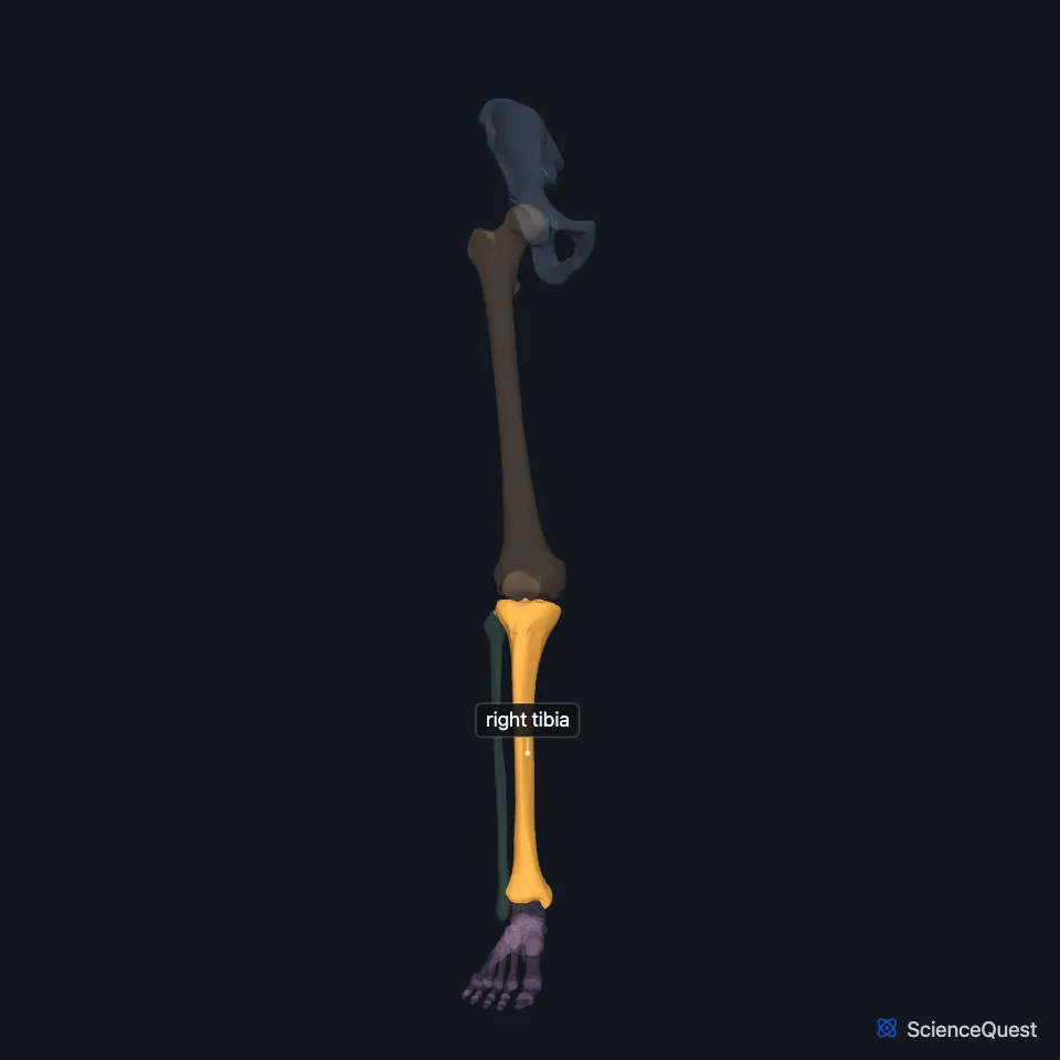
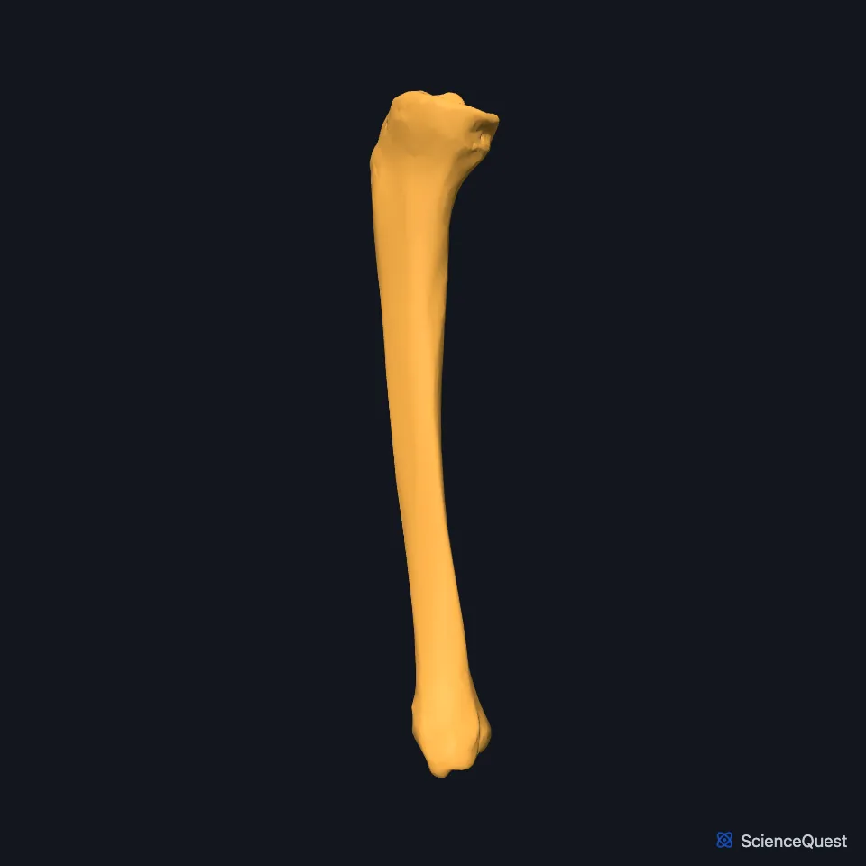
Open it in the Lower Limb Skeleton Explorer See it alone in 3D
Where is the tibia?
The tibia runs down the medial side of the leg, the region between the knee and ankle joints, beside the fibula. The interosseous membrane joins the two bones. (OpenStax A&P 2e, 8.4; StatPearls, Tibia)
Most of its medial surface is covered only by skin, so the shin can be felt all the way down the leg. (OpenStax A&P 2e, 8.4; StatPearls, Tibia)
What are the parts of the tibia?
The wide upper end carries a medial and a lateral condyle with flat tops, the tibial plateaus. Between the condyles rises the intercondylar eminence, and the rough areas in front of and behind it anchor the cruciate ligaments and the menisci. (OpenStax A&P 2e, 8.4; StatPearls, Tibia; Gray’s Anatomy (1918), Tibia, p. 256)
The tibial tuberosity, on the front near the top, is where the tendon from the patella ends. Below it the shaft becomes triangular, and the anterior border runs down from the tuberosity along the bone. (OpenStax A&P 2e, 8.4; StatPearls, Tibia; Gray’s Anatomy (1918), Tibia, p. 256)
On the back, the soleal line runs down and medially across the upper third, and the soleus arises from it. (OpenStax A&P 2e, 8.4; StatPearls, Leg Posterior Compartment)
At the lower end, the medial malleolus forms the bump on the inner side of the ankle. On the lateral side, the fibular notch takes the lower end of the fibula. (OpenStax A&P 2e, 8.4; StatPearls, Tibia; Gray’s Anatomy (1918), Tibiofibular joints, p. 347)
Which joints does the tibia form?
Its condyles meet those of the femur at the medial and lateral tibiofemoral joints of the knee. The medial and lateral menisci sit between them, resting on the edges of the tibial condyles and anchored to the tibia. (OpenStax A&P 2e, 8.4 and 9.6; StatPearls, Tibia; Gray’s Anatomy (1918), Tibia, p. 256)
The anterior and posterior cruciate ligaments anchor to the tibia in the intercondylar area, in front of and behind the eminence, and run up to the femoral condyles. (OpenStax A&P 2e, 9.6; StatPearls, Knee and Tibia)
Its lower end and the inside of the medial malleolus meet the talus at the ankle joint. That part of the joint carries body weight from the leg to the foot. (OpenStax A&P 2e, 8.4 and 9.6; StatPearls, Tibia, Ankle Joint and Foot Talus)
The fibula meets the tibia under its lateral condyle at the proximal tibiofibular joint and in the fibular notch at the distal one. (OpenStax A&P 2e, 8.4; Gray’s Anatomy (1918), Tibia, p. 256 and Tibiofibular joints, p. 347)
What does the tibia do?
Body weight passes from the tibia to the talus and on to the calcaneus, which rests on the ground. StatPearls adds that the medial part of the tibia bears most of the load. (OpenStax A&P 2e, 8.4; StatPearls, Tibia)
Eleven muscles arise from or insert on the tibia. They extend and flex the knee and dorsiflex and plantar flex the ankle. (StatPearls, Tibia)
Where the sources differ
StatPearls’ Tibia article names the soleal line as the origin of the soleus, flexor digitorum longus and tibialis posterior. Its Leg Posterior Compartment article places flexor digitorum longus on the middle third of the back of the tibia, and its Tibialis Posterior Muscle article places that muscle on the upper half of the back of the tibia, the middle of the back of the fibula and the interosseous membrane. (StatPearls, Tibia, Leg Posterior Compartment and Tibialis Posterior Muscle)
| Feature | Detail and source |
|---|---|
| Bone type | Long bone, the second longest in the body (OpenStax A&P 2e, 6.2 and 8.4; Gray’s Anatomy (1918), Classes of bones, p. 79 and Tibia, p. 256) |
| Articulates with | Femur (knee); fibula (proximal and distal tibiofibular joints); talus (ankle) (OpenStax A&P 2e, 8.4; StatPearls, Tibia) |
| Muscles attached | 11 (StatPearls, Tibia) |
Touches in this model
Measured on the BodyParts3D meshes: surfaces within 2 mm of the tibia, nearest first, 12 of 24.
- Right fibula in contact
- Right talus in contact
- Interosseous membrane of right leg in contact
- Medial head of right gastrocnemius in contact
- Right extensor digitorum longus in contact
- Right extensor hallucis longus in contact
- Right fibularis brevis in contact
- Right fibularis tertius in contact
- Right flexor digitorum longus in contact
- Right flexor hallucis longus in contact
- Right iliotibial tract in contact
- Right popliteus in contact
Sources
- OpenStax Anatomy and Physiology 2e, section 8.4, Bones of the Lower Limb. CC BY-NC-SA 4.0. Read 2026-10-25.
- StatPearls, Anatomy, Bony Pelvis and Lower Limb: Tibia. Matthew Bourne; Margaret A. Sinkler; Patrick B. Murphy. Last update August 8, 2023. CC BY-NC-ND 4.0. Read 2026-10-25.
- Gray’s Anatomy of the Human Body, 20th US edition (1918), edited by Warren H. Lewis, Osteology, The Tibia (pp. 256 to 260); 1918 name: intercondyloid eminence for intercondylar eminence. Henry Gray; Warren H. Lewis (editor). Public domain. Read 2026-10-25.
- StatPearls, Anatomy, Bony Pelvis and Lower Limb: Leg Posterior Compartment. Rami Abuqubo; Steven B. Graefe; James La Rose. Last update May 13, 2026. CC BY-NC-ND 4.0. Read 2026-10-25.
- Gray’s Anatomy of the Human Body, 20th US edition (1918), edited by Warren H. Lewis, Syndesmology, Tibiofibular Articulation and Tibiofibular Syndesmosis (pp. 347 to 349). Henry Gray; Warren H. Lewis (editor). Public domain. Read 2026-10-25.
- OpenStax Anatomy and Physiology 2e, section 9.6, Anatomy of Selected Synovial Joints. CC BY-NC-SA 4.0. Read 2026-10-25.
- StatPearls, Anatomy, Bony Pelvis and Lower Limb, Knee. Marco Gupton; Onyebuchi Imonugo; Asa C. Black; Marjorie V. Launico; Robert R. Terreberry. Last update November 5, 2023. CC BY-NC-ND 4.0. Read 2026-10-25.
- StatPearls, Anatomy, Bony Pelvis and Lower Limb: Ankle Joint. Daniel Manganaro; Khalid Alsayouri. Last update May 23, 2023. CC BY-NC-ND 4.0. Read 2026-10-25.
- StatPearls, Anatomy, Bony Pelvis and Lower Limb, Foot Talus. Irfan A. Khan; Matthew A. Varacallo. Last update August 8, 2023. CC BY-NC-ND 4.0. Read 2026-10-25.
- OpenStax Anatomy and Physiology 2e, section 6.2, Bone Classification. CC BY-NC-SA 4.0. Read 2026-10-25.
- Gray’s Anatomy of the Human Body, 20th US edition (1918), edited by Warren H. Lewis, Osteology, introduction: long, short, flat and irregular bones (p. 79). Henry Gray; Warren H. Lewis (editor). Public domain. Read 2026-10-25.
- StatPearls, Anatomy, Bony Pelvis and Lower Limb: Tibialis Posterior Muscle. Nicholas M. Corcoran; Matthew A. Varacallo. Last update August 28, 2023. CC BY-NC-ND 4.0. Read 2026-10-25.
The values are facts and free to use; this page’s selection and presentation are © 2026 ScienceQuest.