Talus
The talus is the highest of the seven tarsal bones. Its upper surface meets the tibia and fibula to form the ankle joint. (OpenStax A&P 2e, 8.4; Gray’s Anatomy (1918), Talus, p. 266; StatPearls, Foot and Ankle and Foot Talus)
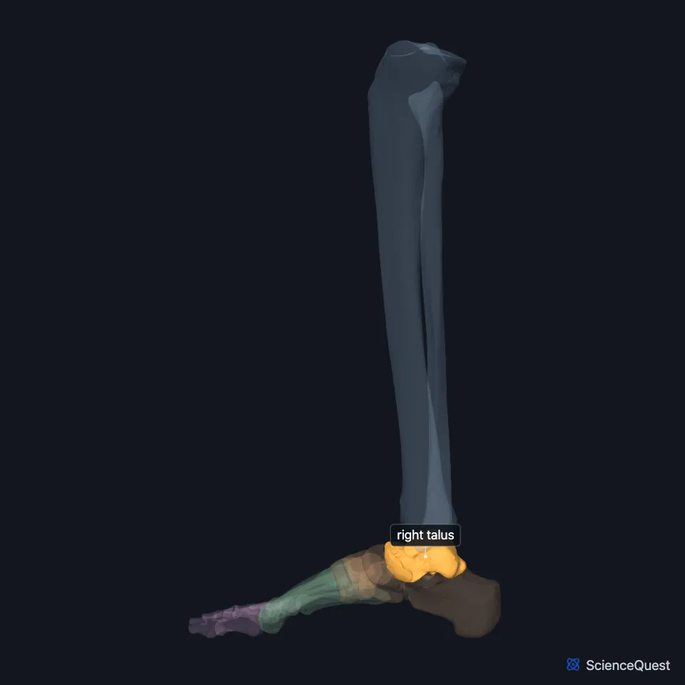
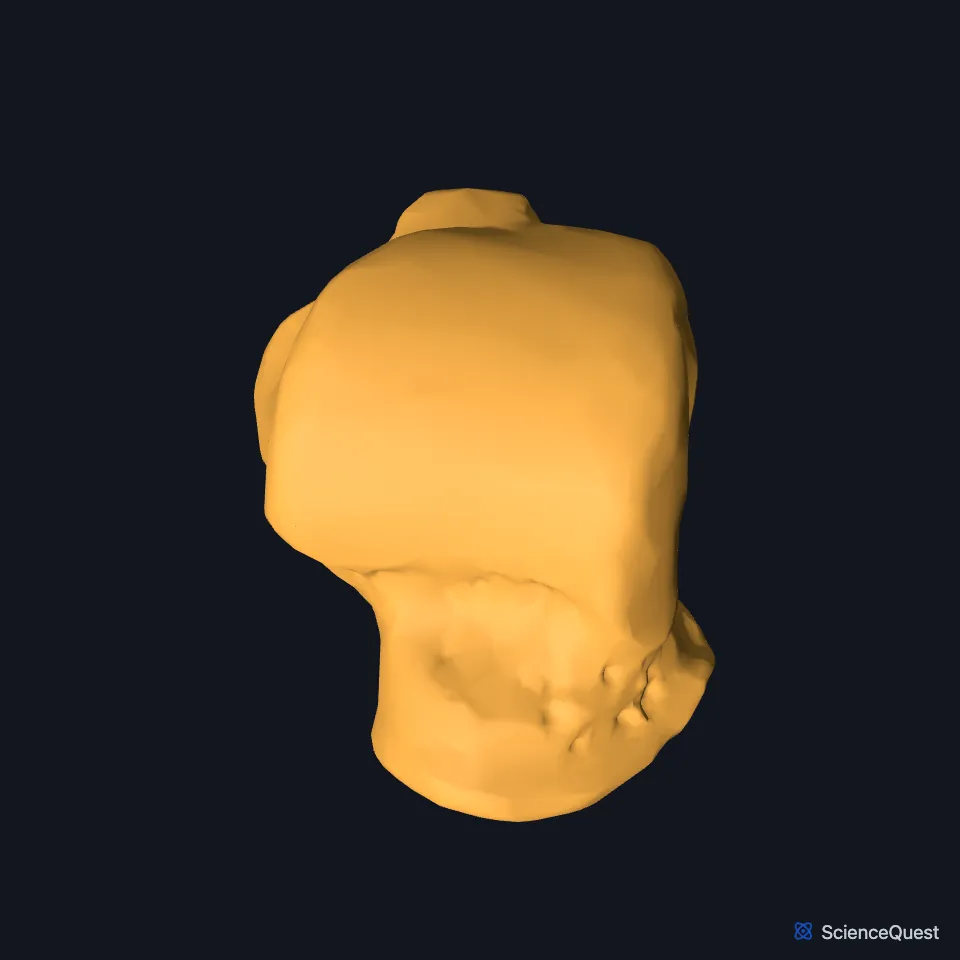
Open it in the Ankle and Foot Skeleton Explorer See it alone in 3D
Where is the talus?
The talus sits at the top of the back half of the foot. The calcaneus lies below it and the navicular bone in front. (OpenStax A&P 2e, 8.4; Gray’s Anatomy (1918), Talus, p. 266; StatPearls, Foot and Ankle)
It sits at the top of the medial longitudinal arch of the foot, receives the weight of the body, and passes it to both longitudinal arches through the subtalar joint. (OpenStax A&P 2e, 8.4; StatPearls, Arches of the Foot and Foot Talus)
What are the parts of the talus?
StatPearls describes the talus as a saddle-shaped bone with a head, a neck and a body. The head points forward to the navicular, and the talar dome on top of the body meets the tibia and fibula. (StatPearls, Foot Talus)
No muscle attaches to the talus, and its blood supply is limited. Ligaments do attach, among them the lateral ankle ligaments and the deltoid ligament complex on the medial side. (StatPearls, Foot Talus)
At its back, the tendon of flexor hallucis longus runs between the medial and lateral tubercles. (StatPearls, Foot Talus)
Which joints does the talus form?
Three surfaces make the ankle joint: the top of the talus meets the lower end of the tibia, its superomedial surface meets the medial malleolus, and its lateral side meets the lateral malleolus of the fibula. (OpenStax A&P 2e, 8.4; StatPearls, Foot Talus and Ankle Joint)
The malleoli grip its sides, so the ankle works as a hinge for dorsiflexion and plantar flexion. The StatPearls ankle chapter also lists inversion and eversion at the ankle; OpenStax and the StatPearls talus chapter give those movements to the joints below the talus. (OpenStax A&P 2e, 9.6; StatPearls, Foot Talus and Ankle Joint)
Below, it meets the calcaneus at the subtalar joint, which turns the sole inward and outward in inversion and eversion of the foot. Its joint with the navicular also contributes to those movements. (OpenStax A&P 2e, 9.6; StatPearls, Foot Talus)
A shelf on the medial side of the calcaneus, the sustentaculum tali, supports the medial side of the talus. (OpenStax A&P 2e, 8.4; StatPearls, Foot Talus; Gray’s Anatomy (1918), Talus, p. 266)
What does the talus do?
The talus is the bridge between leg and foot. Body weight passes from the tibia to the talus and on to the calcaneus, which rests on the ground. (StatPearls, Foot Talus; OpenStax A&P 2e, 8.4)
Through the subtalar joint it also passes body weight to the medial and lateral longitudinal arches, which helps keep the foot aligned. (StatPearls, Foot Talus)
| Feature | Detail and source |
|---|---|
| Bone type | Short bone, the highest of the seven tarsals (OpenStax A&P 2e, 6.2 and 8.4; Gray’s Anatomy (1918), Classes of bones, p. 79 and Talus, p. 266) |
| Articulates with | Tibia and fibula (ankle); calcaneus (subtalar joint); navicular bone (OpenStax A&P 2e, 8.4 and 9.6; StatPearls, Foot Talus) |
| Muscle attachments | None (StatPearls, Foot Talus) |
Touches in this model
Measured on the BodyParts3D meshes: surfaces within 2 mm of the talus, nearest first.
- Navicular bone of right foot in contact
- Right calcaneus in contact
- Right tibia in contact
- Right extensor hallucis brevis in contact
- Right fibularis brevis in contact
- Skin in contact
- Right tibialis posterior 1.4 mm away
- Right fibularis longus 1.5 mm away
- Right flexor hallucis longus 1.6 mm away
Sources
- OpenStax Anatomy and Physiology 2e, section 8.4, Bones of the Lower Limb. CC BY-NC-SA 4.0. Read 2026-10-25.
- Gray’s Anatomy of the Human Body, 20th US edition (1918), edited by Warren H. Lewis, Osteology, The Talus (pp. 266 to 269). Henry Gray; Warren H. Lewis (editor). Public domain. Read 2026-10-25.
- StatPearls, Anatomy, Bony Pelvis and Lower Limb: Foot and Ankle. Jennifer Ficke; Adegbenro O. Fakoya; Doug W. Byerly. Last update May 24, 2026. CC BY-NC-ND 4.0. Read 2026-10-25.
- StatPearls, Anatomy, Bony Pelvis and Lower Limb, Foot Talus. Irfan A. Khan; Matthew A. Varacallo. Last update August 8, 2023. CC BY-NC-ND 4.0. Read 2026-10-25.
- StatPearls, Anatomy, Bony Pelvis and Lower Limb: Arches of the Foot. Hiteshkumar M. Chauhan; Muhammad Taqi. Last update August 27, 2025. CC BY-NC-ND 4.0. Read 2026-10-25.
- StatPearls, Anatomy, Bony Pelvis and Lower Limb: Ankle Joint. Daniel Manganaro; Khalid Alsayouri. Last update May 23, 2023. CC BY-NC-ND 4.0. Read 2026-10-25.
- OpenStax Anatomy and Physiology 2e, section 9.6, Anatomy of Selected Synovial Joints. CC BY-NC-SA 4.0. Read 2026-10-25.
- OpenStax Anatomy and Physiology 2e, section 6.2, Bone Classification. CC BY-NC-SA 4.0. Read 2026-10-25.
- Gray’s Anatomy of the Human Body, 20th US edition (1918), edited by Warren H. Lewis, Osteology, introduction: long, short, flat and irregular bones (p. 79). Henry Gray; Warren H. Lewis (editor). Public domain. Read 2026-10-25.
The values are facts and free to use; this page’s selection and presentation are © 2026 ScienceQuest.