Calcaneus
The calcaneus, or heel bone, is the largest bone of the foot and forms the heel. It is one of the seven tarsal bones. (OpenStax A&P 2e, 8.4; StatPearls, Calcaneus)
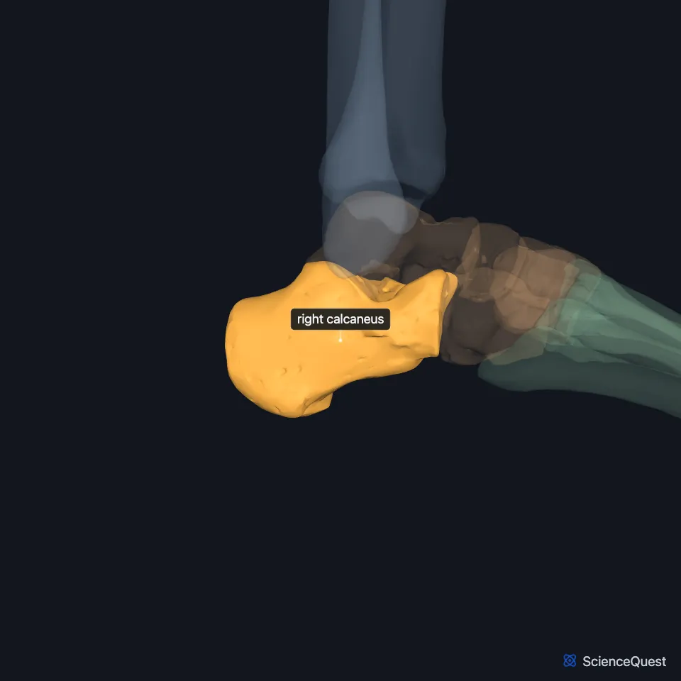
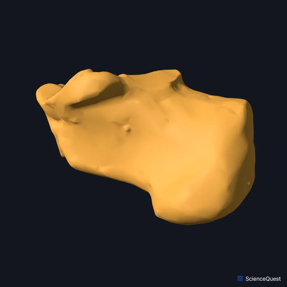
Open it in the Ankle and Foot Skeleton Explorer See it alone in 3D
Where is the calcaneus?
The calcaneus lies in the hindfoot, below the talus and behind the midfoot. Its long axis runs about along the midline of the foot. (StatPearls, Calcaneus)
In front, the cuboid bone articulates with it. (OpenStax A&P 2e, 8.4; StatPearls, Calcaneus; Gray’s Anatomy (1918), Calcaneus, p. 263)
What are the parts of the calcaneus?
Low on its back is the calcaneal tuberosity. On its lateral surface, the fibular trochlea separates the tendon of fibularis brevis, which passes above it, from that of fibularis longus below. (StatPearls, Calcaneus)
On its medial surface, the sustentaculum tali juts out to support the talus, and a groove beneath it carries the tendon of flexor hallucis longus. (OpenStax A&P 2e, 8.4; StatPearls, Calcaneus)
Grooves on the calcaneus and talus meet to form the tarsal sinus, a space between the two bones that holds vessels, nerves, ligaments and fat. (StatPearls, Calcaneus)
Which joints and tendons does the calcaneus have?
Its upper surface carries the anterior and posterior talar facets, and the sustentaculum tali carries the middle one; all three meet the talus. The anterior and middle facets also take part, with the head of the talus and the navicular bone, in the talocalcaneonavicular joint. Its front face carries the facet for the calcaneocuboid joint. (StatPearls, Calcaneus and Navicular Bone)
The gastrocnemius, soleus and plantaris join in the calcaneal (Achilles) tendon, which inserts on the calcaneus. (OpenStax A&P 2e, 11.6; StatPearls, Calcaneus)
The plantar aponeurosis runs from the calcaneus to the toes and keeps the longitudinal arch from collapsing under body weight. On the lateral side of the ankle, the calcaneofibular ligament ties it to the fibula. (OpenStax A&P 2e, 11.6 and 9.6; StatPearls, Foot Fascia and Ankle Joint)
What does the calcaneus do?
Body weight passes from the tibia through the talus to the calcaneus, which rests on the ground. At the back of the longitudinal arches, it is the support where the arches meet the ground. (OpenStax A&P 2e, 8.4; Gray’s Anatomy (1918), Calcaneus, p. 263; StatPearls, Foot Talus and Arches of the Foot)
It anchors the Achilles tendon, through which the gastrocnemius, soleus and plantaris plantar flex the foot in standing, walking, running and jumping. Muscles that move the toes attach to it too. (StatPearls, Calcaneus)
How often does the calcaneus break?
The calcaneus is the tarsal bone that breaks most often: StatPearls gives it 60% of tarsal fractures and names falls from a height and road accidents as the main causes. (StatPearls, Calcaneus)
Rare extra ossicles can form near it: the os sustentaculi behind the sustentaculum tali, and the os calcaneus secundarius between the calcaneus, cuboid, talus and navicular. (StatPearls, Calcaneus)
Where the sources differ
OpenStax gives dorsiflexion and plantar flexion to the ankle joint and inversion and eversion to the subtalar joint. StatPearls lists all four movements for the subtalar joint. (OpenStax A&P 2e, 9.6; StatPearls, Calcaneus)
StatPearls’ Calcaneus article names the joint through all three talar facets the talocalcaneal, or subtalar, joint. Its Navicular Bone article treats the talocalcaneonavicular joint and the posterior talocalcaneal joint as two joints that together form the subtalar articular complex. (StatPearls, Calcaneus and Navicular Bone)
| Feature | Detail and source |
|---|---|
| Bone type | Short bone, the largest tarsal (OpenStax A&P 2e, 6.2; StatPearls, Calcaneus) |
| Articulates with | Talus (subtalar joint); cuboid bone (calcaneocuboid joint) (StatPearls, Calcaneus) |
| Tendon | Calcaneal (Achilles) tendon (OpenStax A&P 2e, 11.6; StatPearls, Achilles Tendon) |
Touches in this model
Measured on the BodyParts3D meshes: surfaces within 2 mm of the calcaneus, nearest first, 12 of 15.
- Right cuboid bone in contact
- Right talus in contact
- Abductor digiti minimi of right foot in contact
- Right abductor hallucis in contact
- Right calcaneal tendon in contact
- Right extensor digitorum brevis in contact
- Right extensor hallucis brevis in contact
- Right fibularis brevis in contact
- Right fibularis longus in contact
- Right flexor accessorius in contact
- Right flexor digitorum brevis in contact
- Right long plantar ligament in contact
Sources
- OpenStax Anatomy and Physiology 2e, section 8.4, Bones of the Lower Limb. CC BY-NC-SA 4.0. Read 2026-10-25.
- StatPearls, Anatomy, Bony Pelvis and Lower Limb: Calcaneus. Marco Gupton; Meltem Özdemir; Robert R. Terreberry. Last update May 23, 2023. CC BY-NC-ND 4.0. Read 2026-10-25.
- Gray’s Anatomy of the Human Body, 20th US edition (1918), edited by Warren H. Lewis, Osteology, The Calcaneus (pp. 263 to 266). Henry Gray; Warren H. Lewis (editor). Public domain. Read 2026-10-25.
- StatPearls, Anatomy, Bony Pelvis and Lower Limb: Navicular Bone. Devindra Prapto; Mark A. Dreyer. Last update August 21, 2023. CC BY-NC-ND 4.0. Read 2026-10-25.
- OpenStax Anatomy and Physiology 2e, section 11.6, Appendicular Muscles of the Pelvic Girdle and Lower Limbs. CC BY-NC-SA 4.0. Read 2026-10-25.
- OpenStax Anatomy and Physiology 2e, section 9.6, Anatomy of Selected Synovial Joints. CC BY-NC-SA 4.0. Read 2026-10-25.
- StatPearls, Anatomy, Bony Pelvis and Lower Limb, Foot Fascia. Matthew Bourne; Aditi Talkad; Matthew A. Varacallo. Last update August 14, 2023. CC BY-NC-ND 4.0. Read 2026-10-25.
- StatPearls, Anatomy, Bony Pelvis and Lower Limb: Ankle Joint. Daniel Manganaro; Khalid Alsayouri. Last update May 23, 2023. CC BY-NC-ND 4.0. Read 2026-10-25.
- StatPearls, Anatomy, Bony Pelvis and Lower Limb, Foot Talus. Irfan A. Khan; Matthew A. Varacallo. Last update August 8, 2023. CC BY-NC-ND 4.0. Read 2026-10-25.
- StatPearls, Anatomy, Bony Pelvis and Lower Limb: Arches of the Foot. Hiteshkumar M. Chauhan; Muhammad Taqi. Last update August 27, 2025. CC BY-NC-ND 4.0. Read 2026-10-25.
- OpenStax Anatomy and Physiology 2e, section 6.2, Bone Classification. CC BY-NC-SA 4.0. Read 2026-10-25.
- StatPearls, Anatomy, Bony Pelvis and Lower Limb: Achilles Tendon. Michael Wong; Achraf H. Jardaly; John Kiel. Last update August 8, 2023. CC BY-NC-ND 4.0. Read 2026-10-25.
The values are facts and free to use; this page’s selection and presentation are © 2026 ScienceQuest.