Fibula
The fibula is the slender bone on the lateral side of the leg. It bears no weight, and the many muscles attached to it shape its shaft. (OpenStax A&P 2e, 8.4; StatPearls, Fibula; Gray’s Anatomy (1918), Fibula, p. 260)
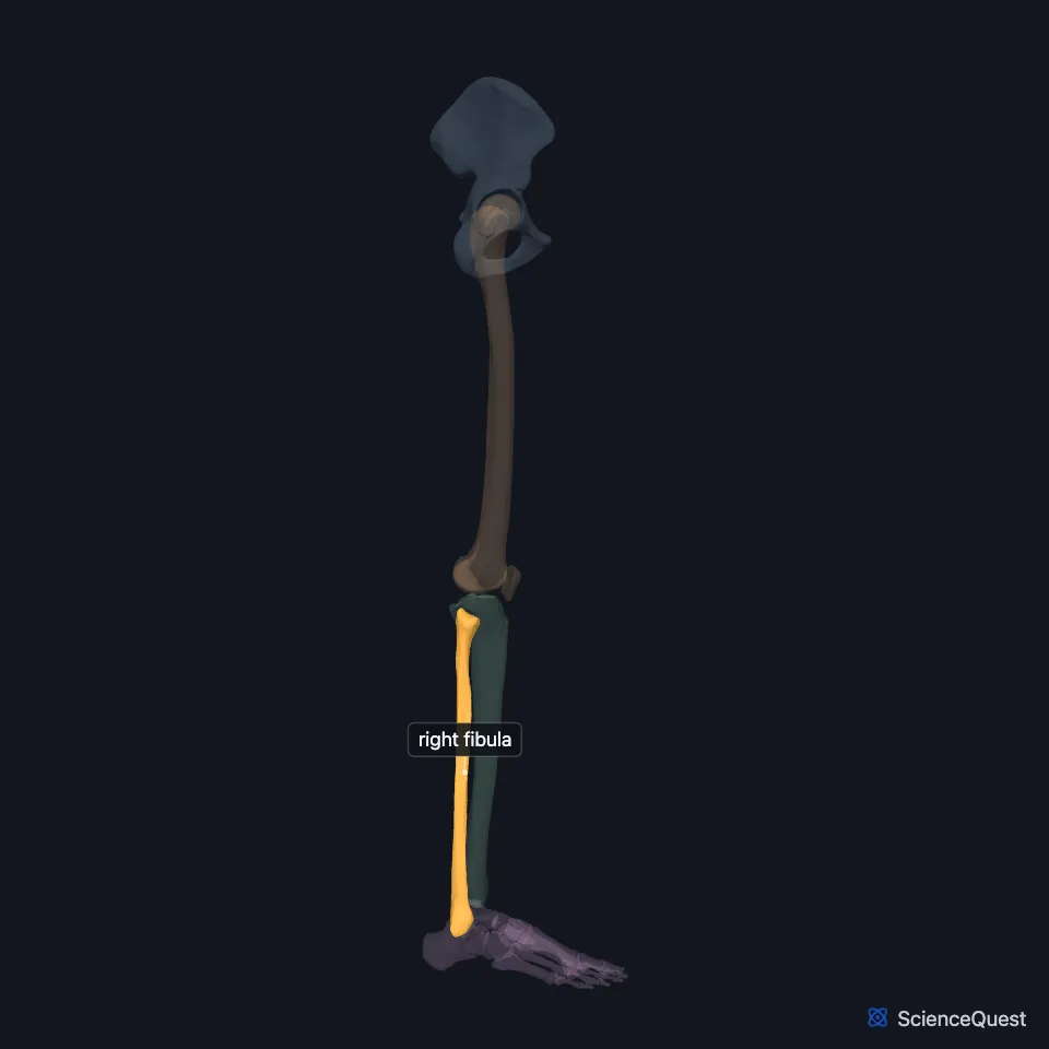
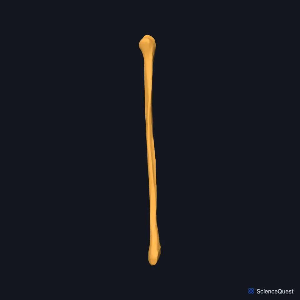
Open it in the Lower Limb Skeleton Explorer See it alone in 3D
Where is the fibula?
The fibula starts just behind the head of the tibia at the knee and runs down the lateral side of the leg to the ankle. StatPearls calls it much smaller and thinner than the tibia. (StatPearls, Fibula)
Muscles cover most of its shaft. Its lower end, the lateral malleolus, stands out at the ankle, where you can feel it under the skin. (OpenStax A&P 2e, 8.4; StatPearls, Fibula; Gray’s Anatomy (1918), Fibula, p. 260)
What are the parts of the fibula?
It has a head, a neck, a shaft and a lower end. The head is the small knob at the top, and the lower end forms the lateral malleolus, the bump on the outer side of the ankle. (StatPearls, Fibula; OpenStax A&P 2e, 8.4)
A narrow ridge down the medial side of the shaft, the interosseous border, anchors the interosseous membrane that spans to the tibia. The shaft’s shape follows its muscles: triangular above and more irregular lower down. (OpenStax A&P 2e, 8.4; StatPearls, Fibula)
Which joints and ligaments does the fibula have?
Its head meets the underside of the lateral condyle of the tibia at the proximal tibiofibular joint, and its lower end sits in the fibular notch of the tibia at the distal one. (OpenStax A&P 2e, 8.4; StatPearls, Fibula; Gray’s Anatomy (1918), Tibiofibular joints, p. 347)
The deep side of the lateral malleolus meets the talus in the ankle joint. With the medial malleolus of the tibia it holds the talus from the sides, and the two malleoli and the lower end of the tibia make the socket of the ankle, the mortise. (OpenStax A&P 2e, 8.4 and 9.6; StatPearls, Fibula and Ankle Joint)
On the outer side of the knee, the fibular collateral ligament joins the head of the fibula to the lateral epicondyle of the femur. At the ankle, three ligaments bind the lower fibula: the anterior and posterior talofibular ligaments to the talus and the calcaneofibular ligament to the calcaneus. (OpenStax A&P 2e, 9.6; StatPearls, Fibula and Ankle Joint; Gray’s Anatomy (1918), Femur, p. 242)
The biceps femoris tendon attaches to the head. The fibularis longus and brevis attach to the lateral side, and the extensor digitorum longus and extensor hallucis longus to the medial side. (StatPearls, Fibula)
The fibularis tertius arises from the lower front of the fibula and the interosseous membrane, and its tendon inserts on the dorsal base of the fifth metatarsal. (OpenStax A&P 2e, 11.6; StatPearls, Leg Anterior Compartment)
What does the fibula do?
With the tibia, the fibula keeps the ankle joint stable. Grooves at its lower end anchor the ligaments that steady the ankle as it moves. (StatPearls, Fibula; OpenStax A&P 2e, 8.4)
Above the ankle, a strong syndesmosis binds the lower ends of the tibia and fibula. Its short interosseous fibers are the chief bond between the two bones, and below them the talus sits in the pocket the two malleoli make. (OpenStax A&P 2e, 9.6; Gray’s Anatomy (1918), Tibiofibular joints, p. 347; StatPearls, Foot Talus)
Does the fibula vary between people?
The fibularis tertius is not present in everyone; StatPearls says it can vary in size or be absent 20% of the time. A fibularis quartus muscle appears in the lateral compartment in about 12% to 22%. (OpenStax A&P 2e, 11.6; StatPearls, Fibula)
Where the sources differ
OpenStax says the fibula serves mainly for muscle attachment. StatPearls says its main function is to combine with the tibia to stabilize the ankle joint. (OpenStax A&P 2e, 8.4; StatPearls, Fibula)
| Feature | Detail and source |
|---|---|
| Bone type | Long bone (OpenStax A&P 2e, 6.2; StatPearls, Fibula; Gray’s Anatomy (1918), Classes of bones, p. 79) |
| Articulates with | Tibia (proximal and distal tibiofibular joints); talus (ankle) (OpenStax A&P 2e, 8.4; StatPearls, Fibula; Gray’s Anatomy (1918), Tibiofibular joints, p. 347) |
| Weight bearing | None (OpenStax A&P 2e, 8.4; StatPearls, Fibula) |
Touches in this model
Measured on the BodyParts3D meshes: surfaces within 2 mm of the fibula, nearest first, 12 of 13.
- Right tibia in contact
- Long head of right biceps femoris in contact
- Right extensor digitorum longus in contact
- Right fibularis brevis in contact
- Right fibularis longus in contact
- Right flexor hallucis longus in contact
- Right soleus in contact
- Short head of right biceps femoris in contact
- Skin in contact
- Interosseous membrane of right leg 0.7 mm away
- Right extensor hallucis longus 1.2 mm away
- Lateral head of right gastrocnemius 1.5 mm away
Sources
- OpenStax Anatomy and Physiology 2e, section 8.4, Bones of the Lower Limb. CC BY-NC-SA 4.0. Read 2026-10-25.
- StatPearls, Anatomy, Bony Pelvis and Lower Limb: Fibula. Marco Gupton; Akul Munjal; Michael Kang. Last update May 23, 2023. CC BY-NC-ND 4.0. Read 2026-10-25.
- Gray’s Anatomy of the Human Body, 20th US edition (1918), edited by Warren H. Lewis, Osteology, The Fibula (pp. 260 to 263). Henry Gray; Warren H. Lewis (editor). Public domain. Read 2026-10-25.
- Gray’s Anatomy of the Human Body, 20th US edition (1918), edited by Warren H. Lewis, Syndesmology, Tibiofibular Articulation and Tibiofibular Syndesmosis (pp. 347 to 349). Henry Gray; Warren H. Lewis (editor). Public domain. Read 2026-10-25.
- OpenStax Anatomy and Physiology 2e, section 9.6, Anatomy of Selected Synovial Joints. CC BY-NC-SA 4.0. Read 2026-10-25.
- StatPearls, Anatomy, Bony Pelvis and Lower Limb: Ankle Joint. Daniel Manganaro; Khalid Alsayouri. Last update May 23, 2023. CC BY-NC-ND 4.0. Read 2026-10-25.
- Gray’s Anatomy of the Human Body, 20th US edition (1918), edited by Warren H. Lewis, Osteology, The Femur (pp. 242 to 255). Henry Gray; Warren H. Lewis (editor). Public domain. Read 2026-10-25.
- OpenStax Anatomy and Physiology 2e, section 11.6, Appendicular Muscles of the Pelvic Girdle and Lower Limbs. CC BY-NC-SA 4.0. Read 2026-10-25.
- StatPearls, Anatomy, Bony Pelvis and Lower Limb: Leg Anterior Compartment. Bradley Lezak; Marjorie V. Launico. Last update June 8, 2026. CC BY-NC-ND 4.0. Read 2026-10-25.
- StatPearls, Anatomy, Bony Pelvis and Lower Limb, Foot Talus. Irfan A. Khan; Matthew A. Varacallo. Last update August 8, 2023. CC BY-NC-ND 4.0. Read 2026-10-25.
- OpenStax Anatomy and Physiology 2e, section 6.2, Bone Classification. CC BY-NC-SA 4.0. Read 2026-10-25.
- Gray’s Anatomy of the Human Body, 20th US edition (1918), edited by Warren H. Lewis, Osteology, introduction: long, short, flat and irregular bones (p. 79). Henry Gray; Warren H. Lewis (editor). Public domain. Read 2026-10-25.
The values are facts and free to use; this page’s selection and presentation are © 2026 ScienceQuest.