Soleus
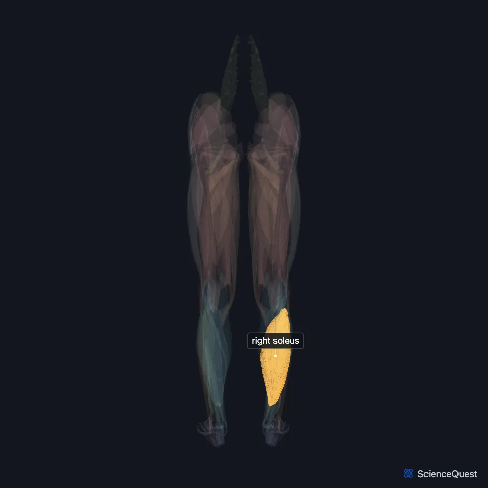
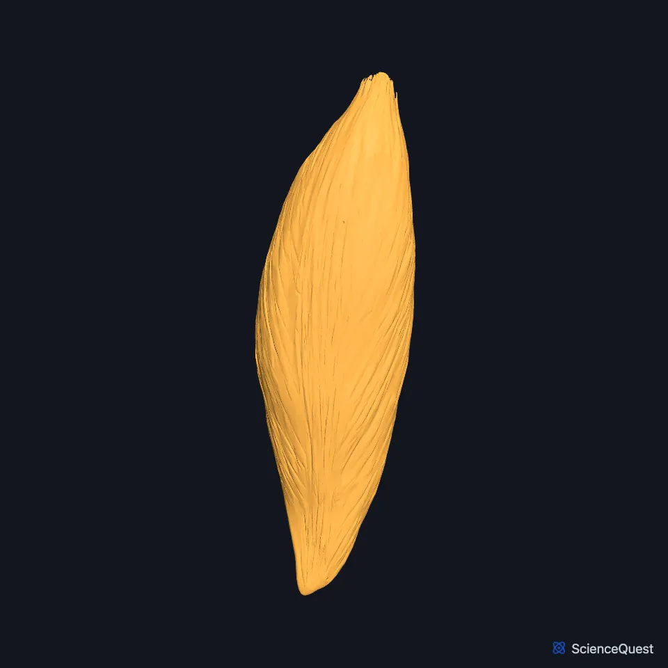
The soleus is a wide, flat muscle of the calf that lies deep to the gastrocnemius. The two join in the calcaneal tendon, which inserts on the back of the heel. (OpenStax A&P 2e, 11.6; StatPearls, Calf; Gray’s Anatomy (1918), Posterior Crural Muscles)
Open it in the Lower Limb Muscles Explorer See it alone in 3D
Where is the soleus?
It sits in the superficial layer of the posterior compartment of the leg, with the gastrocnemius and the plantaris. The transverse intermuscular septum separates this layer from the deep muscles. (StatPearls, Leg Posterior Compartment)
It arises from the back of the head and upper shaft of the fibula and from the soleal line on the back of the tibia. (StatPearls, Leg Posterior Compartment)
How is the soleus built?
A fibrous arch between the head of the fibula and the soleal line adds to its origin. The tibial nerve and the posterior tibial artery pass through this tendinous arch on their way down the leg. (StatPearls, Gastrocnemius Muscle and Tibial Nerve)
Its aponeurosis joins that of the gastrocnemius in the lower third of the leg, and the calcaneal tendon carries both to the tuberosity of the calcaneus. (StatPearls, Gastrocnemius Muscle)
Unlike the gastrocnemius, it crosses only one joint. (StatPearls, Gastrocnemius Muscle)
What does the soleus do?
It plantarflexes the ankle whatever the position of the knee. (StatPearls, Leg Posterior Compartment)
It contributes most of the plantarflexion in walking, though not in running. With more aerobic, red fibers than the gastrocnemius, it carries sustained work such as standing and walking uphill. (StatPearls, Gastrocnemius Muscle and Calf)
The calf muscles are large and powerful, and you use them all the time to stand and walk. When you stand, the soleus steadies the leg on the foot and keeps you from tipping forward. (OpenStax A&P 2e, 11.6; Gray’s Anatomy (1918), Posterior Crural Muscles)
What supplies the soleus?
The tibial nerve passes through the popliteal fossa and gives branches to the soleus, the gastrocnemius, the popliteus and the plantaris. (StatPearls, Calf)
The sural arteries, branches of the popliteal artery, supply it along with the heads of the gastrocnemius. (StatPearls, Gastrocnemius Muscle)
The popliteus, the highest of the deep muscles of the compartment, inserts on the back of the tibia just above the origin of the soleus. (StatPearls, Leg Posterior Compartment)
Does the soleus vary?
Its fiber arrangement, shape and tendon vary between people. One cadaver study sorted it into bipennate, unipennate, multipennate and nonpennate patterns, and some people have an accessory soleus. (StatPearls, Calf)
Where the sources differ
Sources give different root levels for the tibial nerve: S1 and S2 for its supply to the calf muscles in the StatPearls calf article, L4 to S3 for the nerve as a whole in its tibial nerve article. (StatPearls, Calf and Tibial Nerve)
| Feature | Detail and source |
|---|---|
| Origin | Back of the fibular head and upper fibula; soleal line of the tibia; fibrous arch between them (StatPearls, Gastrocnemius Muscle) |
| Insertion | Tuberosity of the calcaneus, through the calcaneal tendon (StatPearls, Gastrocnemius Muscle) |
| Action | Plantarflexes the ankle at any knee position (StatPearls, Leg Posterior Compartment) |
| Nerve | Tibial nerve (StatPearls, Calf) |
Touches in this model
Measured on the BodyParts3D meshes: surfaces within 2 mm of the soleus, nearest first, 12 of 14.
- Lateral head of right gastrocnemius in contact
- Long head of right biceps femoris in contact
- Medial head of right gastrocnemius in contact
- Right calcaneal tendon in contact
- Right fibularis brevis in contact
- Right fibularis longus in contact
- Right flexor digitorum longus in contact
- Right flexor hallucis longus in contact
- Right plantaris in contact
- Right popliteus in contact
- Right tibialis posterior in contact
- Right fibula in contact
Sources
- OpenStax Anatomy and Physiology 2e, section 11.6, Appendicular Muscles of the Pelvic Girdle and Lower Limbs. CC BY-NC-SA 4.0. Read 2026-10-25.
- StatPearls, Anatomy, Bony Pelvis and Lower Limb: Calf. Justin T. Binstead; Matthew A. Varacallo. Last update January 31, 2026. CC BY-NC-ND 4.0. Read 2026-10-25.
- Anatomy of the Human Body (Gray’s Anatomy), 20th US edition, 1918, Myology, The Muscles and Fasciae of the Leg, 2. The Posterior Crural Muscles, superficial group, pages 482 to 485; Internet Archive scan of the 1918 Lea and Febiger printing, terminology of 1918. Henry Gray; edited by Warren H. Lewis. Public domain. Read 2026-10-25.
- StatPearls, Anatomy, Bony Pelvis and Lower Limb: Leg Posterior Compartment. Rami Abuqubo; Steven B. Graefe; James La Rose. Last update May 13, 2026. CC BY-NC-ND 4.0. Read 2026-10-25.
- StatPearls, Anatomy, Bony Pelvis and Lower Limb: Gastrocnemius Muscle. Bruno Bordoni; Matthew A. Varacallo. Last update April 11, 2026. CC BY-NC-ND 4.0. Read 2026-10-25.
- StatPearls, Anatomy, Bony Pelvis and Lower Limb: Tibial Nerve. Sohil S. Desai; James La Rose. Last update September 14, 2026. CC BY-NC-ND 4.0. Read 2026-10-25.
The values are facts and free to use; this page’s selection and presentation are © 2026 ScienceQuest.