Navicular bone of foot
The navicular bone of the foot is a wedge-shaped tarsal bone of the midfoot. It sits between the head of the talus behind and the three cuneiform bones in front. (StatPearls, Navicular Bone; OpenStax A&P 2e, 8.4)
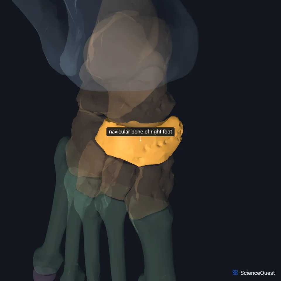
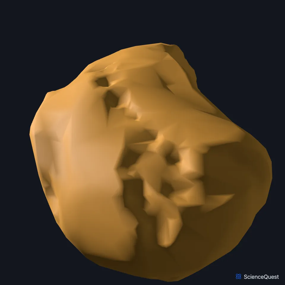
Open it in the Ankle and Foot Skeleton Explorer See it alone in 3D
Where is the navicular bone?
It lies in the midfoot with the cuboid and the three cuneiforms, and it articulates with five tarsal bones: the talus, the cuboid and the medial, intermediate and lateral cuneiforms. (StatPearls, Navicular Bone; OpenStax A&P 2e, 8.4)
What are the parts of the navicular bone?
Its back is concave and covered in cartilage, and it takes the head of the talus. Its front divides into three facets: the largest for the medial cuneiform, a flat one for the intermediate and the smallest for the lateral. (StatPearls, Navicular Bone)
On its medial end, the navicular tuberosity takes the tendon of tibialis posterior and the plantar and medial navicular ligaments. (StatPearls, Navicular Bone)
Which tendons and joints hold the navicular bone?
Tibialis posterior is the only tendon that inserts on the navicular. StatPearls names it the main dynamic stabilizer of the hindfoot, because it preserves the medial longitudinal arch. (StatPearls, Navicular Bone)
With the head of the talus and the anterior and middle articular surfaces of the calcaneus, it forms the talocalcaneonavicular joint, called the acetabulum pedis for its likeness to the hip. The plantar calcaneonavicular (spring) ligament supports that joint on the medial side. (StatPearls, Navicular Bone)
What does the navicular bone do?
Its three joints with the cuneiforms converge underneath and form the transverse tarsal arch of the foot. (StatPearls, Navicular Bone)
Its joint with the talus takes part in inversion and eversion of the foot, alongside the subtalar joint. (OpenStax A&P 2e, 9.6; StatPearls, Foot Talus)
Does the navicular bone vary between people?
Some people have an accessory navicular, an extra ossicle beside the navicular tuberosity that makes a bony bump on the medial side of the foot. (StatPearls, Navicular Bone)
Where the sources differ
OpenStax classes every joint between tarsal bones as a plane joint. StatPearls describes the navicular’s joint with the head of the talus as a ball-and-socket joint. (OpenStax A&P 2e, 9.6; StatPearls, Navicular Bone)
| Feature | Detail and source |
|---|---|
| Bone type | Short bone, one of the seven tarsals (OpenStax A&P 2e, 6.2 and 8.4; Gray’s Anatomy (1918), Classes of bones, p. 79; StatPearls, Foot and Ankle) |
| Articulates with | Talus, cuboid and the three cuneiforms (StatPearls, Navicular Bone) |
| Tendon | Tibialis posterior, the only one (StatPearls, Navicular Bone) |
Touches in this model
Measured on the BodyParts3D meshes: surfaces within 2 mm of the navicular bone of foot, nearest first.
- Right cuboid bone in contact
- Right intermediate cuneiform bone in contact
- Right talus in contact
- Right extensor hallucis brevis in contact
- Right tibialis posterior in contact
- Skin in contact
- Right medial cuneiform bone 0.2 mm away
- Right lateral cuneiform bone 0.6 mm away
- Right tibialis anterior 1.0 mm away
Sources
- StatPearls, Anatomy, Bony Pelvis and Lower Limb: Navicular Bone. Devindra Prapto; Mark A. Dreyer. Last update August 21, 2023. CC BY-NC-ND 4.0. Read 2026-10-25.
- OpenStax Anatomy and Physiology 2e, section 8.4, Bones of the Lower Limb. CC BY-NC-SA 4.0. Read 2026-10-25.
- OpenStax Anatomy and Physiology 2e, section 9.6, Anatomy of Selected Synovial Joints. CC BY-NC-SA 4.0. Read 2026-10-25.
- StatPearls, Anatomy, Bony Pelvis and Lower Limb, Foot Talus. Irfan A. Khan; Matthew A. Varacallo. Last update August 8, 2023. CC BY-NC-ND 4.0. Read 2026-10-25.
- OpenStax Anatomy and Physiology 2e, section 6.2, Bone Classification. CC BY-NC-SA 4.0. Read 2026-10-25.
- Gray’s Anatomy of the Human Body, 20th US edition (1918), edited by Warren H. Lewis, Osteology, introduction: long, short, flat and irregular bones (p. 79). Henry Gray; Warren H. Lewis (editor). Public domain. Read 2026-10-25.
- StatPearls, Anatomy, Bony Pelvis and Lower Limb: Foot and Ankle. Jennifer Ficke; Adegbenro O. Fakoya; Doug W. Byerly. Last update May 24, 2026. CC BY-NC-ND 4.0. Read 2026-10-25.
The values are facts and free to use; this page’s selection and presentation are © 2026 ScienceQuest.