Patella
The patella, or kneecap, is the largest sesamoid bone in the body. It sits in the tendon of the quadriceps femoris, in front of the knee joint. (OpenStax A&P 2e, 8.4; StatPearls, Knee Patella)
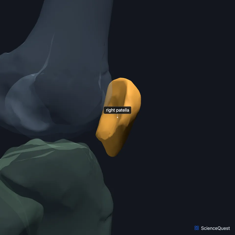
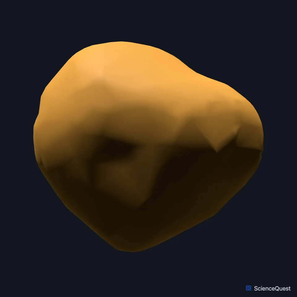
Where is the patella?
The patella lies in front of the knee joint, under the fascia lata and the tendon of rectus femoris. Below it, the patellar ligament runs on to the tibial tuberosity on the front of the tibia. (StatPearls, Knee Patella; OpenStax A&P 2e, 11.6 and 8.4)
What is a sesamoid bone?
A sesamoid bone sits inside the tendon of a muscle where the tendon crosses a joint. Sesamoid bones vary in number and position from person to person, and the patella is present in everyone. (OpenStax A&P 2e, 8.4 and 6.2; StatPearls, Sesamoid Bones)
Which bone and muscles does the patella meet?
The patella articulates with one bone, the femur, at the patellar surface on its lower end. It does not articulate with the tibia. (OpenStax A&P 2e, 8.4; Gray’s Anatomy (1918), Patella, p. 255; StatPearls, Knee Patella)
At the femoropatellar joint it slides up and down in a groove on the femur. That joint shares one articular capsule with the two tibiofemoral joints of the knee. (OpenStax A&P 2e, 9.6; Gray’s Anatomy (1918), Knee joint, p. 339; StatPearls, Knee)
Four muscles attach to it: the rectus femoris and vastus intermedius to its upper third, and the vastus medialis and vastus lateralis to its medial and lateral borders. (StatPearls, Knee Patella)
What does the patella do?
By keeping the quadriceps tendon off the femur, the patella protects it from friction. It also lengthens the moment arm of the patellar ligament, which makes the quadriceps a stronger extensor of the knee. (OpenStax A&P 2e, 8.4; StatPearls, Knee Patella and Sesamoid Bones)
Through the patella and the patellar ligament, the quadriceps femoris extends the leg at the knee. The patella also shields the deeper structures of the joint. (OpenStax A&P 2e, 9.6; StatPearls, Knee Patella)
The last 15 degrees of extension need twice the torque of the first. By lengthening the moment arm, the patella adds 60% more torque over that range. (StatPearls, Knee Patella)
Why does the patella pull to the side?
As the knee straightens, the patella ends up a little to the lateral side of the groove on the femur. The depth of that groove and the height of its lateral wall help keep the patella in line, and a flat lateral side of the groove, one form of trochlear dysplasia, is a common cause of recurrent patellar instability. A Q-angle of 20° or more tends to pull the patella’s tracking to the lateral side. (OpenStax A&P 2e, 8.4; StatPearls, Knee Patella and Medial Patellofemoral Ligament)
Patellofemoral syndrome, or runner’s knee, is among the most common causes of pain at the front of the knee. Overuse is its main contributing factor, and it affects women about twice as often as men. (OpenStax A&P 2e, 8.4; StatPearls, Patellofemoral Syndrome)
Where the sources differ
The sources disagree on which sesamoid bones everyone has. OpenStax says the patellae are the only sesamoid bones common to every person, while Gray’s Anatomy (1918) also calls two at the metacarpophalangeal joint of the thumb constant and two at the metatarsophalangeal joint of the great toe always present. (OpenStax A&P 2e, 6.2; Gray’s Anatomy (1918), The Sesamoid Bones)
| Feature | Detail and source |
|---|---|
| Bone type | Sesamoid bone, the largest in the body (OpenStax A&P 2e, 8.4; StatPearls, Knee Patella and Sesamoid Bones) |
| Lies in | Tendon of the quadriceps femoris (OpenStax A&P 2e, 8.4; StatPearls, Knee Patella; Gray’s Anatomy (1918), Patella, p. 255) |
| Articulates with | Femur (patellar surface) only (OpenStax A&P 2e, 8.4; Gray’s Anatomy (1918), Patella, p. 255) |
Touches in this model
Measured on the BodyParts3D meshes: surfaces within 2 mm of the patella, nearest first.
- Right rectus femoris in contact
- Right vastus intermedius in contact
- Right vastus medialis in contact
- Skin in contact
- Right iliotibial tract 0.1 mm away
- Right vastus lateralis 0.5 mm away
- Right femur 1.7 mm away
Sources
- OpenStax Anatomy and Physiology 2e, section 8.4, Bones of the Lower Limb. CC BY-NC-SA 4.0. Read 2026-10-25.
- StatPearls, Anatomy, Bony Pelvis and Lower Limb, Knee Patella. Chandler F. Cox; Margaret A. Sinkler; Asa C. Black; Marjorie V. Launico; John B. Hubbard. Last update October 27, 2023. CC BY-NC-ND 4.0. Read 2026-10-25.
- OpenStax Anatomy and Physiology 2e, section 11.6, Appendicular Muscles of the Pelvic Girdle and Lower Limbs. CC BY-NC-SA 4.0. Read 2026-10-25.
- OpenStax Anatomy and Physiology 2e, section 6.2, Bone Classification. CC BY-NC-SA 4.0. Read 2026-10-25.
- StatPearls, Anatomy, Sesamoid Bones. Anthony Y. Yeung; Tafline C. Arbor; Dr. Rohin Garg. Last update April 4, 2023. CC BY-NC-ND 4.0. Read 2026-10-25.
- Gray’s Anatomy of the Human Body, 20th US edition (1918), edited by Warren H. Lewis, Osteology, The Patella (pp. 255 to 256). Henry Gray; Warren H. Lewis (editor). Public domain. Read 2026-10-25.
- OpenStax Anatomy and Physiology 2e, section 9.6, Anatomy of Selected Synovial Joints. CC BY-NC-SA 4.0. Read 2026-10-25.
- Gray’s Anatomy of the Human Body, 20th US edition (1918), edited by Warren H. Lewis, Syndesmology, The Knee-joint (pp. 339 to 347). Henry Gray; Warren H. Lewis (editor). Public domain. Read 2026-10-25.
- StatPearls, Anatomy, Bony Pelvis and Lower Limb, Knee. Marco Gupton; Onyebuchi Imonugo; Asa C. Black; Marjorie V. Launico; Robert R. Terreberry. Last update November 5, 2023. CC BY-NC-ND 4.0. Read 2026-10-25.
- StatPearls, Anatomy, Bony Pelvis and Lower Limb: Medial Patellofemoral Ligament. Dylan Luxenburg; Michael G. Rizzo. Last update April 17, 2023. CC BY-NC-ND 4.0. Read 2026-10-25.
- StatPearls, Patellofemoral Syndrome. Jared M. Bump; Lindsay Lewis. Last update February 13, 2023. CC BY-NC-ND 4.0. Read 2026-10-25.
- Anatomy of the Human Body (Gray’s Anatomy), 20th US edition, 1918, Osteology, The Sesamoid Bones (Ossa Sesamoidea), pages 277 to 278; Internet Archive scan of the 1918 Lea and Febiger printing, terminology of 1918. Henry Gray; edited by Warren H. Lewis. Public domain. Read 2026-10-25.
The values are facts and free to use; this page’s selection and presentation are © 2026 ScienceQuest.