Vastus lateralis
The vastus lateralis is the largest of the four muscles of the quadriceps femoris, and it lies on the outer side of the thigh. (StatPearls, Vastus Lateralis Muscle; OpenStax A&P 2e, 11.6)
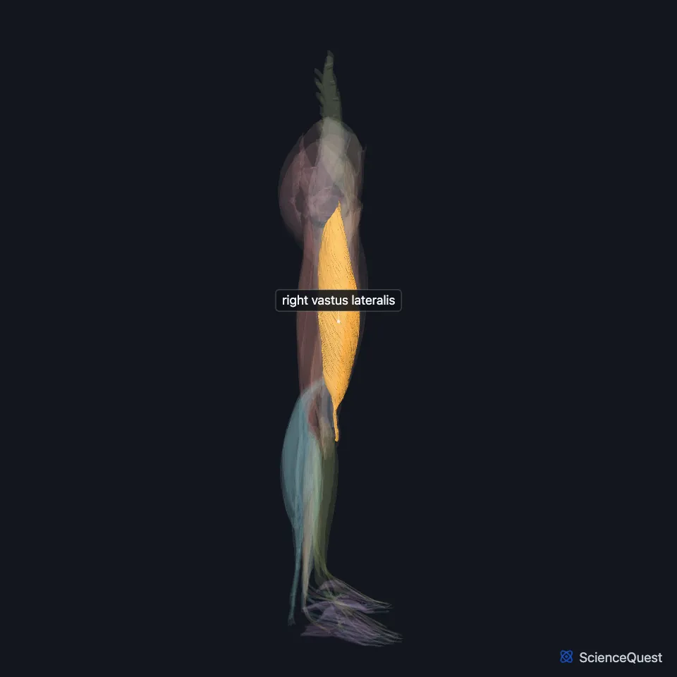
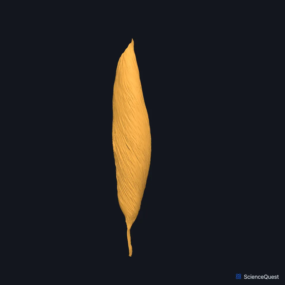
Open it in the Lower Limb Muscles Explorer See it alone in 3D
Where is the vastus lateralis?
It lies on the outer side of the femur inside the fascia lata, which thickens over it into the iliotibial tract. (StatPearls, Vastus Lateralis Muscle)
At the level of the greater trochanter, rectus femoris forms its front inner border. The lateral intermuscular septum separates it from the long and short heads of biceps femoris in the back compartment. (StatPearls, Vastus Lateralis Muscle)
The deep fibers of the gluteus maximus insert on the gluteal tuberosity between vastus lateralis and adductor magnus. (StatPearls, Gluteus Maximus Muscle)
Its name gives its side: it covers the outer side of the femur, and the vastus medialis covers the inner side. (OpenStax A&P 2e, 11.2; Gray’s Anatomy (1918), Anterior Femoral Muscles)
Where does the vastus lateralis attach?
It has a broad, continuous origin on the upper femur: the intertrochanteric line, the greater trochanter, the gluteal tuberosity, the lateral lip of the linea aspera and the lateral intermuscular septum. (StatPearls, Vastus Lateralis Muscle)
Its fibers join the quadriceps tendon, attach to the outer side of the patella, and end on the tibial tuberosity through the patellar ligament. (StatPearls, Vastus Lateralis Muscle; OpenStax A&P 2e, 11.6)
What does the vastus lateralis do?
It is a primary extensor of the knee, and with vastus medialis it stabilizes the knee joint. (StatPearls, Vastus Lateralis Muscle)
Its force grows as the knee bends further. With the knee straight it exerts a small force that holds the position with little effort. (StatPearls, Thigh Quadriceps Muscle)
Near the knee its fibers combine with those of vastus medialis in the patellar retinaculum, which helps keep the patella aligned over the femur. (StatPearls, Vastus Lateralis Muscle)
What supplies the vastus lateralis?
Muscular branches of the femoral nerve enter it, carrying fibers from L2 to L4, with L3 the main root. (StatPearls, Vastus Lateralis Muscle)
The lateral circumflex femoral artery is its main supply. Perforating branches of the deep femoral artery add to it, piercing the lateral intermuscular septum to reach the front compartment. (StatPearls, Vastus Lateralis Muscle)
Does the vastus lateralis vary?
About 60% of specimens show two insertional heads, a long head and a vastus lateralis obliquus, usually parted by a layer of fat or fascia. (StatPearls, Vastus Lateralis Muscle)
| Feature | Detail and source |
|---|---|
| Origin | Intertrochanteric line, greater trochanter, gluteal tuberosity, lateral lip of the linea aspera, lateral intermuscular septum (StatPearls, Vastus Lateralis Muscle) |
| Insertion | Outer side of the patella through the quadriceps tendon; tibial tuberosity through the patellar ligament (StatPearls, Vastus Lateralis Muscle; OpenStax A&P 2e, 11.6) |
| Action | Extends the knee; with vastus medialis, stabilizes it (StatPearls, Vastus Lateralis Muscle) |
| Nerve | Femoral nerve (L2 to L4, mainly L3) (StatPearls, Vastus Lateralis Muscle) |
Touches in this model
Measured on the BodyParts3D meshes: surfaces within 2 mm of the vastus lateralis, nearest first.
- Long head of right biceps femoris in contact
- Right gluteus maximus in contact
- Right iliotibial tract in contact
- Right rectus femoris in contact
- Right vastus intermedius in contact
- Right vastus medialis in contact
- Short head of right biceps femoris in contact
- Right femur in contact
- Right tibia in contact
- Skin in contact
- Right patella 0.5 mm away
- Right adductor magnus 1.3 mm away
Sources
- StatPearls, Anatomy, Bony Pelvis and Lower Limb: Vastus Lateralis Muscle. Nicholas L. Biondi; Matthew A. Varacallo. Last update August 8, 2023. CC BY-NC-ND 4.0. Read 2026-10-25.
- OpenStax Anatomy and Physiology 2e, section 11.6, Appendicular Muscles of the Pelvic Girdle and Lower Limbs. CC BY-NC-SA 4.0. Read 2026-10-25.
- StatPearls, Anatomy, Bony Pelvis and Lower Limb, Gluteus Maximus Muscle. Adel Elzanie; Judith Borger. Last update April 1, 2023. CC BY-NC-ND 4.0. Read 2026-10-25.
- OpenStax Anatomy and Physiology 2e, section 11.2, Naming Skeletal Muscles. CC BY-NC-SA 4.0. Read 2026-10-25.
- Anatomy of the Human Body (Gray’s Anatomy), 20th US edition, 1918, Myology, The Muscles and Fasciae of the Thigh, 1. The Anterior Femoral Muscles, pages 467 to 471; Internet Archive scan of the 1918 Lea and Febiger printing, terminology of 1918. Henry Gray; edited by Warren H. Lewis. Public domain. Read 2026-10-25.
- StatPearls, Anatomy, Bony Pelvis and Lower Limb: Thigh Quadriceps Muscle. Bruno Bordoni; Matthew A. Varacallo. Last update May 8, 2023. CC BY-NC-ND 4.0. Read 2026-10-25.
The values are facts and free to use; this page’s selection and presentation are © 2026 ScienceQuest.