Vastus medialis
The vastus medialis is the quadriceps femoris muscle on the inner side of the thigh. Like the other vasti, it extends the knee and steadies the patella. (OpenStax A&P 2e, 11.6; StatPearls, Anterior Thigh Muscles; Gray’s Anatomy (1918), Anterior Femoral Muscles)
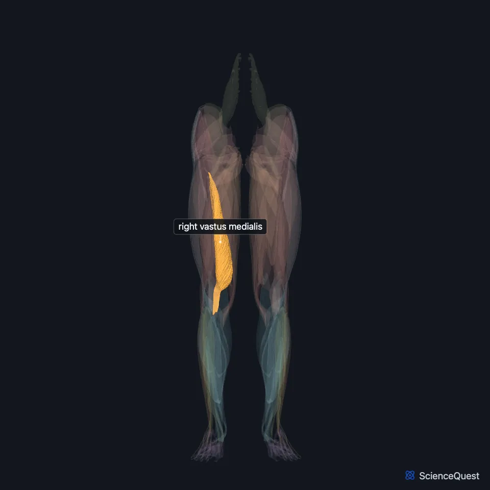
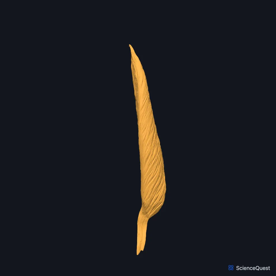
Open it in the Lower Limb Muscles Explorer See it alone in 3D
Where is the vastus medialis?
It lies on the inner side of the thigh. Rectus femoris runs down the front and vastus lateralis down the outer side, and vastus intermedius lies in front of the femur under rectus femoris. Only when rectus femoris is lifted away does a narrow gap show between vastus intermedius and vastus medialis. (OpenStax A&P 2e, 11.6; Gray’s Anatomy (1918), Anterior Femoral Muscles)
It forms the front outer wall of the adductor canal, the passage deep to sartorius through the middle third of the thigh that carries the femoral artery, the femoral vein and the saphenous nerve. (StatPearls, Adductor Canal, Medial Thigh Muscles and Anterior Thigh Muscles)
Where does the vastus medialis attach?
It arises along the medial lip of the linea aspera on the back of the femur, and higher up from the lower part of the intertrochanteric line. (StatPearls, Anterior Thigh Muscles)
It inserts on the inner base and border of the patella, forming the medial patellar retinaculum and the inner side of the quadriceps tendon. (StatPearls, Anterior Thigh Muscles)
What does the vastus medialis do?
Its own insertion on the patellar tendon is small and cannot hold the patella medially as the knee extends. Instead it pulls on the aponeurosis of vastus intermedius, which counters the outward pull of vastus lateralis on the patella. (StatPearls, Thigh Quadriceps Muscle)
With vastus lateralis it stabilizes the knee joint. (StatPearls, Vastus Lateralis Muscle)
The quadriceps femoris extends the leg at the knee. Below the patella its common tendon carries on as the patellar ligament to the tuberosity of the tibia. (OpenStax A&P 2e, 9.6; Gray’s Anatomy (1918), Anterior Femoral Muscles and The Knee-joint)
What supplies the vastus medialis?
Its nerve is one of the four motor branches of the posterior division of the femoral nerve, and it follows the muscle’s front inner border. The same nerves to the vasti also supply the knee joint. (StatPearls, Thigh Femoral Nerve and Thigh Quadriceps Muscle)
The other quadriceps muscles take blood from the lateral circumflex femoral artery. The vastus medialis is the exception, supplied by the femoral artery, the deep femoral artery and the superior medial genicular branch of the popliteal artery. (StatPearls, Thigh Muscles)
Is the vastus medialis one muscle or two?
Some authors divide it by fiber direction into a vastus medialis obliquus and a vastus medialis longus, and find more nerve supply in the oblique part. (StatPearls, Thigh Quadriceps Muscle)
Where the sources differ
Sources differ on the upper end of its origin. The StatPearls anterior thigh article gives the lower part of the intertrochanteric line; its quadriceps article gives the anatomical neck of the femur. Both name the medial lip of the linea aspera. (StatPearls, Anterior Thigh Muscles and Thigh Quadriceps Muscle)
The StatPearls anterior thigh article lists the nerve to vastus medialis among the contents of the adductor canal. Its adductor canal article notes that some authors challenge this. (StatPearls, Anterior Thigh Muscles and Adductor Canal)
| Feature | Detail and source |
|---|---|
| Origin | Medial lip of the linea aspera; above it the intertrochanteric line or the femoral neck, by source (StatPearls, Anterior Thigh Muscles and Thigh Quadriceps Muscle) |
| Insertion | Inner base and border of the patella; inner side of the quadriceps tendon (StatPearls, Anterior Thigh Muscles) |
| Action | Extends the knee; steadies the patella (StatPearls, Anterior Thigh Muscles) |
| Nerve | Femoral nerve, posterior division (StatPearls, Thigh Femoral Nerve) |
Touches in this model
Measured on the BodyParts3D meshes: surfaces within 2 mm of the vastus medialis, nearest first, 12 of 14.
- Right adductor brevis in contact
- Right adductor longus in contact
- Right adductor magnus in contact
- Right pectineus in contact
- Right rectus femoris in contact
- Right sartorius in contact
- Right vastus intermedius in contact
- Right vastus lateralis in contact
- Right femur in contact
- Right patella in contact
- Right tibia in contact
- Skin in contact
Sources
- OpenStax Anatomy and Physiology 2e, section 11.6, Appendicular Muscles of the Pelvic Girdle and Lower Limbs. CC BY-NC-SA 4.0. Read 2026-10-25.
- StatPearls, Anatomy, Bony Pelvis and Lower Limb: Anterior Thigh Muscles. Ayesha Khan; Abdul Arain. Last update March 17, 2023. CC BY-NC-ND 4.0. Read 2026-10-25.
- Anatomy of the Human Body (Gray’s Anatomy), 20th US edition, 1918, Myology, The Muscles and Fasciae of the Thigh, 1. The Anterior Femoral Muscles, pages 467 to 471; Internet Archive scan of the 1918 Lea and Febiger printing, terminology of 1918. Henry Gray; edited by Warren H. Lewis. Public domain. Read 2026-10-25.
- StatPearls, Anatomy, Abdomen and Pelvis: Adductor Canal (Subsartorial Canal, Hunter Canal). Allan Migirov; Tafline C. Arbor; Renato C. Vilella. Last update January 9, 2024. CC BY-NC-ND 4.0. Read 2026-10-25.
- StatPearls, Anatomy, Bony Pelvis and Lower Limb: Medial Thigh Muscles. Jordan L. Ramage; Matthew A. Varacallo. Last update August 21, 2023. CC BY-NC-ND 4.0. Read 2026-10-25.
- StatPearls, Anatomy, Bony Pelvis and Lower Limb: Thigh Quadriceps Muscle. Bruno Bordoni; Matthew A. Varacallo. Last update May 8, 2023. CC BY-NC-ND 4.0. Read 2026-10-25.
- StatPearls, Anatomy, Bony Pelvis and Lower Limb: Vastus Lateralis Muscle. Nicholas L. Biondi; Matthew A. Varacallo. Last update August 8, 2023. CC BY-NC-ND 4.0. Read 2026-10-25.
- OpenStax Anatomy and Physiology 2e, section 9.6, Anatomy of Selected Synovial Joints. CC BY-NC-SA 4.0. Read 2026-10-25.
- Anatomy of the Human Body (Gray’s Anatomy), 20th US edition, 1918, Syndesmology, The Knee-joint (Articulatio Genu), the Ligamentum Patellae, page 340; Internet Archive scan of the 1918 Lea and Febiger printing, terminology of 1918. Henry Gray; edited by Warren H. Lewis. Public domain. Read 2026-10-25.
- StatPearls, Anatomy, Bony Pelvis and Lower Limb: Thigh Femoral Nerve. Nader A. Refai; Asa C. Black; Prasanna Tadi. Last update August 22, 2023. CC BY-NC-ND 4.0. Read 2026-10-25.
- StatPearls, Anatomy, Bony Pelvis and Lower Limb: Thigh Muscles. Basem Attum; Matthew A. Varacallo. Last update December 17, 2022. CC BY-NC-ND 4.0. Read 2026-10-25.
The values are facts and free to use; this page’s selection and presentation are © 2026 ScienceQuest.