Rectus femoris
The rectus femoris is the most superficial of the four quadriceps femoris muscles at the front of the thigh. It is the one quadriceps muscle that crosses two joints, so it acts at both the hip and the knee. (StatPearls, Rectus Femoris Muscle; OpenStax A&P 2e, 11.6; Gray’s Anatomy (1918), Anterior Femoral Muscles)
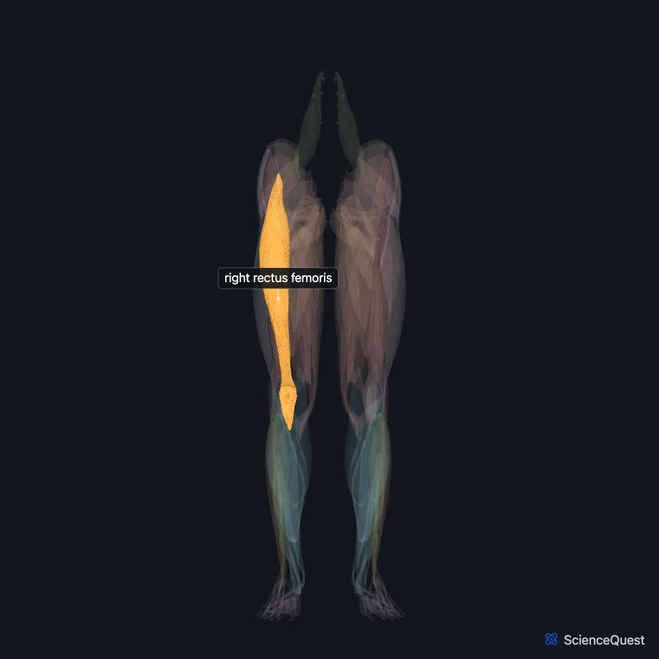
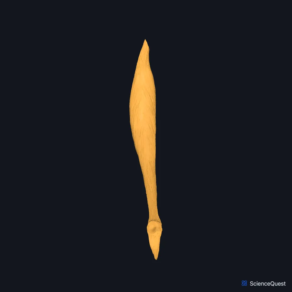
Open it in the Lower Limb Muscles Explorer See it alone in 3D
Where is the rectus femoris?
It runs almost vertically down the front of the thigh. Vastus lateralis lies on the outer side, vastus medialis on the inner side, and vastus intermedius between them, deep to the rectus femoris. (StatPearls, Rectus Femoris Muscle; OpenStax A&P 2e, 11.6; Gray’s Anatomy (1918), Anterior Femoral Muscles)
The rectus femoris takes its name from its straight course down the middle of the front of the thigh. (OpenStax A&P 2e, 11.2 and 11.1; Gray’s Anatomy (1918), Anterior Femoral Muscles)
Where does the rectus femoris attach?
It has two heads. The direct head arises from the anterior inferior iliac spine and the indirect head from the ridge above the acetabulum, and they fuse into a common tendon. (StatPearls, Rectus Femoris Muscle)
Below, it inserts with the other quadriceps muscles on the base of the patella through the quadriceps tendon. Through the patellar ligament the group reaches a second insertion, on the tibial tuberosity. (StatPearls, Rectus Femoris Muscle)
What does the rectus femoris do?
It flexes the hip with sartorius and the iliopsoas, and extends the knee as part of the quadriceps femoris. At both joints it opposes the hamstrings. (StatPearls, Rectus Femoris Muscle)
Because it spans two joints, full extension of the knee limits its pull on the hip, and the reverse. With the hip flexed, as when you sit, it is a weak knee extensor. (StatPearls, Rectus Femoris Muscle)
Its part in forceful knee extension has earned it the name kicking muscle. (StatPearls, Rectus Femoris Muscle)
What supplies the rectus femoris?
The femoral nerve supplies all four parts of the quadriceps femoris, the rectus femoris among them. Its branch enters the rectus femoris near the anterior inferior iliac spine. (StatPearls, Rectus Femoris Muscle and Thigh Quadriceps Muscle)
The lateral circumflex femoral artery supplies the quadriceps. It branches from the deep femoral artery, the largest branch of the femoral artery, though its origin varies a good deal between people. (StatPearls, Rectus Femoris Muscle and Anterior Thigh Muscles)
The patella develops inside the quadriceps tendon. It shields the front of the knee joint and makes the quadriceps pull at a greater angle, which gives it more leverage. (OpenStax A&P 2e, 9.6; Gray’s Anatomy (1918), The Patella)
Where the sources differ
Sources differ on which muscle is the most superficial at the front of the thigh. The StatPearls rectus femoris article gives the rectus femoris; its sartorius and anterior thigh articles give the sartorius, which crosses over the quadriceps. (StatPearls, Rectus Femoris Muscle, Thigh Sartorius Muscle and Anterior Thigh Muscles)
| Feature | Detail and source |
|---|---|
| Origin | Anterior inferior iliac spine (direct head); ridge above the acetabulum (indirect head) (StatPearls, Rectus Femoris Muscle) |
| Insertion | Base of the patella through the quadriceps tendon; tibial tuberosity through the patellar ligament (StatPearls, Rectus Femoris Muscle) |
| Action | Flexes the hip; extends the knee (StatPearls, Rectus Femoris Muscle) |
| Nerve | Femoral nerve (StatPearls, Rectus Femoris Muscle) |
Touches in this model
Measured on the BodyParts3D meshes: surfaces within 2 mm of the rectus femoris, nearest first, 12 of 14.
- Right gluteus minimus in contact
- Right iliacus in contact
- Right iliotibial tract in contact
- Right pectineus in contact
- Right sartorius in contact
- Right tensor fasciae latae in contact
- Right vastus intermedius in contact
- Right vastus lateralis in contact
- Right vastus medialis in contact
- Right hip bone in contact
- Right patella in contact
- Right tibia in contact
Sources
- StatPearls, Anatomy, Abdomen and Pelvis, Rectus Femoris Muscle. Christopher J. Murdock; Andrew Mudreac; Kofi Agyeman. Last update November 13, 2023. CC BY-NC-ND 4.0. Read 2026-10-25.
- OpenStax Anatomy and Physiology 2e, section 11.6, Appendicular Muscles of the Pelvic Girdle and Lower Limbs. CC BY-NC-SA 4.0. Read 2026-10-25.
- Anatomy of the Human Body (Gray’s Anatomy), 20th US edition, 1918, Myology, The Muscles and Fasciae of the Thigh, 1. The Anterior Femoral Muscles, pages 467 to 471; Internet Archive scan of the 1918 Lea and Febiger printing, terminology of 1918. Henry Gray; edited by Warren H. Lewis. Public domain. Read 2026-10-25.
- OpenStax Anatomy and Physiology 2e, section 11.2, Naming Skeletal Muscles. CC BY-NC-SA 4.0. Read 2026-10-25.
- OpenStax Anatomy and Physiology 2e, section 11.1, Interactions of Skeletal Muscles, Their Fascicle Arrangement, and Their Lever Systems. CC BY-NC-SA 4.0. Read 2026-10-25.
- StatPearls, Anatomy, Bony Pelvis and Lower Limb: Thigh Quadriceps Muscle. Bruno Bordoni; Matthew A. Varacallo. Last update May 8, 2023. CC BY-NC-ND 4.0. Read 2026-10-25.
- StatPearls, Anatomy, Bony Pelvis and Lower Limb: Anterior Thigh Muscles. Ayesha Khan; Abdul Arain. Last update March 17, 2023. CC BY-NC-ND 4.0. Read 2026-10-25.
- OpenStax Anatomy and Physiology 2e, section 9.6, Anatomy of Selected Synovial Joints. CC BY-NC-SA 4.0. Read 2026-10-25.
- Anatomy of the Human Body (Gray’s Anatomy), 20th US edition, 1918, Osteology, The Patella (Knee Cap), page 255; Internet Archive scan of the 1918 Lea and Febiger printing, terminology of 1918. Henry Gray; edited by Warren H. Lewis. Public domain. Read 2026-10-25.
- StatPearls, Anatomy, Bony Pelvis and Lower Limb: Thigh Sartorius Muscle. Benjamin B. Walters; Matthew A. Varacallo. Last update August 28, 2023. CC BY-NC-ND 4.0. Read 2026-10-25.
The values are facts and free to use; this page’s selection and presentation are © 2026 ScienceQuest.