Sartorius
The sartorius is the longest muscle in the body, a band-like muscle that crosses the front of the thigh from the pelvis to the inner side of the knee. (StatPearls, Thigh Sartorius Muscle; OpenStax A&P 2e, 11.6; Gray’s Anatomy (1918), Anterior Femoral Muscles)
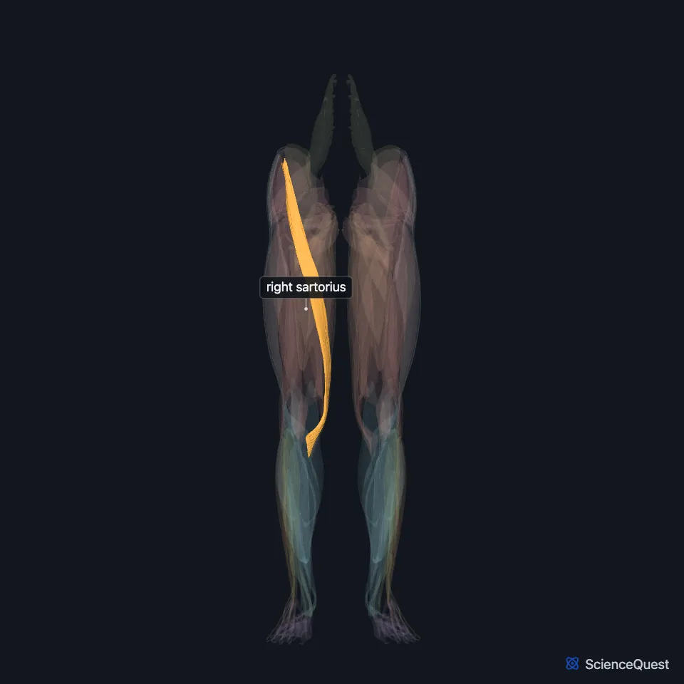
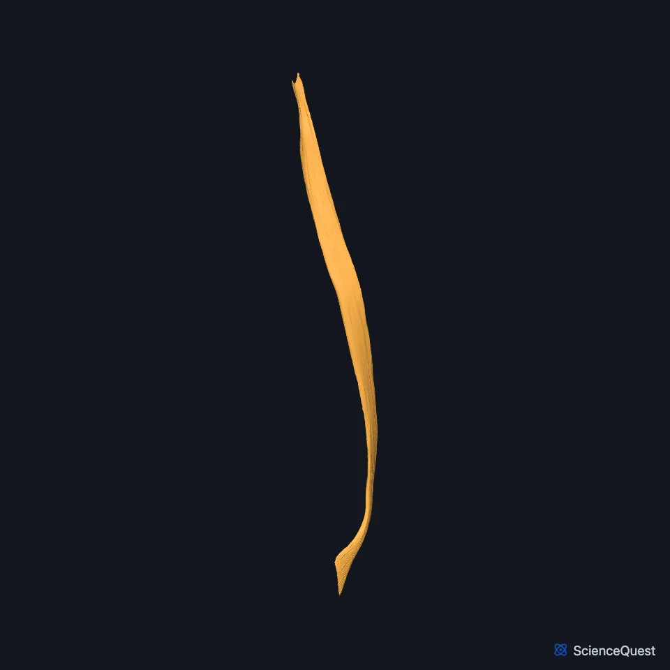
Open it in the Lower Limb Muscles Explorer See it alone in 3D
Where is the sartorius?
It is the most superficial muscle of the front compartment of the thigh. It runs obliquely from the anterior superior iliac spine to the inner side of the upper tibia. (StatPearls, Thigh Sartorius Muscle)
Its long, parallel fibers cross over the quadriceps from the outer side to the inner side. (StatPearls, Anterior Thigh Muscles)
Its name comes from the Latin sartor, a tailor, after the way a tailor placed the legs to work. Working with other hip and knee muscles, it brings the leg into that figure-4 position. (StatPearls, Thigh Sartorius Muscle; OpenStax A&P 2e, 11.6)
Where does the sartorius attach?
It shares its origin at the anterior superior iliac spine with tensor fasciae latae. (StatPearls, Thigh Sartorius Muscle)
On the upper inner tibia its tendon joins those of gracilis and semitendinosus in the pes anserinus, named for its likeness to a goose’s foot. (StatPearls, Thigh Sartorius Muscle and Medial Thigh Muscles)
What does the sartorius do?
At the hip it flexes, abducts and laterally rotates the thigh. At the knee it flexes the leg. (OpenStax A&P 2e, 11.6; Gray’s Anatomy (1918), Anterior Femoral Muscles)
At the knee it also rotates the leg medially, with semimembranosus, semitendinosus, gracilis and popliteus. The two heads of biceps femoris oppose them. (StatPearls, Thigh Sartorius Muscle)
Working with these muscles, it brings the leg into the figure-4 position, the way a tailor sits. (StatPearls, Thigh Sartorius Muscle)
What does the sartorius border?
It is the lateral border of the femoral triangle, with adductor longus on the medial side and the inguinal ligament above. The triangle holds the femoral nerve, artery and vein. (StatPearls, Thigh Femoral Nerve)
Lower down it lies over the adductor canal, superficial to the vastoadductor membrane that roofs the canal. (StatPearls, Adductor Canal)
The anterior division of the femoral nerve supplies it; the femoral nerve carries fibers from L2, L3 and L4. (StatPearls, Thigh Sartorius Muscle)
Muscular branches of the femoral artery provide over half of its blood, with collateral flow from other arteries. (StatPearls, Thigh Sartorius Muscle)
Where the sources differ
Sources differ on which muscle is the most superficial at the front of the thigh. The StatPearls sartorius and anterior thigh articles give the sartorius, which crosses over the quadriceps; its rectus femoris article gives the rectus femoris. (StatPearls, Thigh Sartorius Muscle, Anterior Thigh Muscles and Rectus Femoris Muscle)
| Feature | Detail and source |
|---|---|
| Origin | Anterior superior iliac spine (StatPearls, Thigh Sartorius Muscle) |
| Insertion | Upper inner surface of the tibia, in the pes anserinus (StatPearls, Thigh Sartorius Muscle) |
| Action | Flexes, abducts and laterally rotates the thigh at the hip; flexes the leg at the knee (OpenStax A&P 2e, 11.6; Gray’s Anatomy (1918), Anterior Femoral Muscles) |
| Nerve | Femoral nerve, anterior division (StatPearls, Thigh Sartorius Muscle) |
Touches in this model
Measured on the BodyParts3D meshes: surfaces within 2 mm of the sartorius, nearest first, 12 of 14.
- Right adductor longus in contact
- Right gracilis in contact
- Right iliacus in contact
- Right rectus femoris in contact
- Right tensor fasciae latae in contact
- Right vastus medialis in contact
- Right external oblique in contact
- Right hip bone in contact
- Right inguinal ligament in contact
- Right internal oblique in contact
- Right transversus abdominis in contact
- Skin in contact
Sources
- StatPearls, Anatomy, Bony Pelvis and Lower Limb: Thigh Sartorius Muscle. Benjamin B. Walters; Matthew A. Varacallo. Last update August 28, 2023. CC BY-NC-ND 4.0. Read 2026-10-25.
- OpenStax Anatomy and Physiology 2e, section 11.6, Appendicular Muscles of the Pelvic Girdle and Lower Limbs. CC BY-NC-SA 4.0. Read 2026-10-25.
- Anatomy of the Human Body (Gray’s Anatomy), 20th US edition, 1918, Myology, The Muscles and Fasciae of the Thigh, 1. The Anterior Femoral Muscles, pages 467 to 471; Internet Archive scan of the 1918 Lea and Febiger printing, terminology of 1918. Henry Gray; edited by Warren H. Lewis. Public domain. Read 2026-10-25.
- StatPearls, Anatomy, Bony Pelvis and Lower Limb: Anterior Thigh Muscles. Ayesha Khan; Abdul Arain. Last update March 17, 2023. CC BY-NC-ND 4.0. Read 2026-10-25.
- StatPearls, Anatomy, Bony Pelvis and Lower Limb: Medial Thigh Muscles. Jordan L. Ramage; Matthew A. Varacallo. Last update August 21, 2023. CC BY-NC-ND 4.0. Read 2026-10-25.
- StatPearls, Anatomy, Bony Pelvis and Lower Limb: Thigh Femoral Nerve. Nader A. Refai; Asa C. Black; Prasanna Tadi. Last update August 22, 2023. CC BY-NC-ND 4.0. Read 2026-10-25.
- StatPearls, Anatomy, Abdomen and Pelvis: Adductor Canal (Subsartorial Canal, Hunter Canal). Allan Migirov; Tafline C. Arbor; Renato C. Vilella. Last update January 9, 2024. CC BY-NC-ND 4.0. Read 2026-10-25.
- StatPearls, Anatomy, Abdomen and Pelvis, Rectus Femoris Muscle. Christopher J. Murdock; Andrew Mudreac; Kofi Agyeman. Last update November 13, 2023. CC BY-NC-ND 4.0. Read 2026-10-25.
The values are facts and free to use; this page’s selection and presentation are © 2026 ScienceQuest.