Abdominal Muscles Explorer
Turn the abdominal wall in 3D layer by layer, from rectus abdominis out to transversus abdominis, with the pelvic floor beneath it.
Visualiser
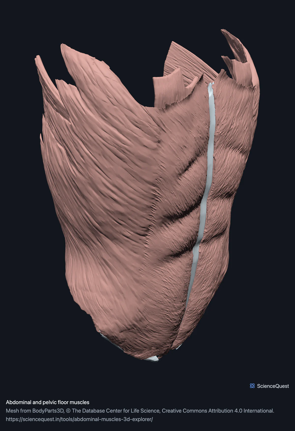
Abdominal and pelvic floor muscles, 25 structures
Checking this device for a stored copy.
Start here
- Hide the wall one layer at a time External oblique, then internal oblique, then transversus abdominis. The fibres run down and forwards in the external oblique, up and forwards in the internal oblique and horizontally in transversus abdominis, which is the arrangement that makes the wall strong and is nearly impossible to read from a picture.
- Click Pelvic floor twice, then Below Opening a band frames it, and this one starts open, so the second click is the one that takes you there. The sling from underneath, which is the only view where puborectalis and the rest of levator ani read as a continuous floor rather than as separate strips.
- Select the linea alba The midline seam where both sides meet. It is where a midline incision goes and where a hernia comes through, and it is tissue rather than muscle.
Muscle, with the linea alba, both inguinal ligaments and the tendinous arch of levator ani. No rectus sheath, which is the most significant absence: the recti arrive as bellies without the aponeurotic wrapping. No peritoneum, no inguinal canal contents and no abdominal organs, which are a separate model.
- 25 structures
- 2888k triangles
- 14000 KB, on request
- 0.00356 mm worst error per axis
- 12 abdominal wall
- 9 pelvic floor
- 4 aponeurosis and ligament
Where this comes from
Segmented from imaging of one adult male body, not drawn by an artist, so the shapes are measurements. Two consequences worth knowing: it is one person’s anatomy rather than an average, and faint stepping on some surfaces is the resolution of the original scan rather than a rendering fault.
Meshes from BodyParts3D, © The Database Center for Life Science, under Creative Commons Attribution 4.0 International. Names and identifiers are the dataset’s own: Foundational Model of Anatomy identifiers and English labels, as its parts list gives them. Converted meshes are shared under the same licence.
Changes made: Meshes were converted from binary STL to an indexed mesh with 16-bit quantised vertex positions, and surface normals were recomputed from the geometry. No vertex was moved other than by that quantisation, whose worst-case error on any one coordinate is reported on the page.
Latin terms for 17 of the 25 structures come from English Wikipedia anatomy infoboxes, under Creative Commons Attribution-ShareAlike 4.0, joined by Wikidata property P1402, Foundational Model of Anatomy identifier. Only a term a source states for that structure is used, never one constructed from a family name, which is why the rest carry no Latin rather than a guess.
All 25 structures
Every structure the model contains, group by group and alphabetically within each. Each name opens the viewer with that structure selected, so a link can point at one structure rather than at the whole region.
Abdominal wall 12
- left external oblique musculus obliquus externus abdominis muscle
- left internal oblique musculus obliquus internus abdominis muscle
- left pyramidalis musculus pyramidalis muscle
- left quadratus lumborum musculus quadratus lumborum muscle
- left rectus abdominis musculus rectus abdominis muscle
- left transversus abdominis musculus transversus abdominis muscle
- right external oblique musculus obliquus externus abdominis muscle
- right internal oblique musculus obliquus internus abdominis muscle
- right pyramidalis musculus pyramidalis muscle
- right quadratus lumborum musculus quadratus lumborum muscle
- right rectus abdominis musculus rectus abdominis muscle
- right transversus abdominis musculus transversus abdominis muscle
Pelvic floor 9
- external anal sphincter sphincter ani externus muscle
- left coccygeus musculus coccygeus muscle
- left iliococcygeus muscle
- left pubococcygeus muscle
- left puborectalis muscle
- right coccygeus musculus coccygeus muscle
- right iliococcygeus muscle
- right pubococcygeus muscle
- right puborectalis muscle
Aponeurosis and ligament 4
- left inguinal ligament ligamentum inguinale other tissue
- linea alba other tissue
- right inguinal ligament ligamentum inguinale other tissue
- tendinous arch of levator ani other tissue
Citing this tool
Last updated . Add the date you accessed it as well, which a citation of a page that can change asks for. If a specific view matters, cite the page address as it stands in your browser instead: it records what is selected and shown.
A wall of sheets, and the seams that hold it
Four of the 25 structures are not muscle, and counting them as muscle would misstate what this is. The linea alba is the midline seam, the inguinal ligaments are the rolled lower border of the external oblique, and the tendinous arch of levator ani is where the pelvic floor hangs from the pelvic wall. That leaves 21 muscles within the 25 structures.
They are here because an abdominal wall without its seams is a set of loose sheets. The seam is also where the clinically interesting things happen.
The floor belongs with the wall
Pubococcygeus, iliococcygeus, puborectalis and coccygeus form the sling the abdominal contents rest on, and the pressure the wall generates is what that sling resists. Separating them would leave both halves of the mechanism incomplete.
Three of the pelvic floor meshes, both pubococcygei and the external anal sphincter, also appear in the reproductive model, because the pelvic floor genuinely belongs to both regions. That is the same overlap the skeleton has with the skull, and it is deliberate rather than duplication.
What the rectus sheath’s absence costs
The rectus abdominis is a belly with no sheath around it, so the model cannot show the sheath’s layers or where their composition changes below the arcuate line. The linea alba is present, which is where the two sheets of the sheath meet in the midline, so the seam is there without the sheet that forms it.
Common questions
Why is the linea alba counted separately?
Because it is not muscle, and pretending otherwise would misstate the count. The linea alba is the midline seam where the aponeuroses of both sides meet, and along with the inguinal ligaments and the tendinous arch of levator ani it is tagged as other tissue, so the model holds 21 muscles within 25 structures. All four are in the model because an abdominal wall without its seams is a set of loose sheets, and the seam is where a midline incision goes and where a hernia comes through.
Can I see the layers separately?
That is what this model is for. External oblique, internal oblique and transversus abdominis are three separate sheets with fibres running in three different directions, which is the arrangement that makes the wall strong, and it is nearly impossible to read from a flat picture. Hide them one at a time from the outside in and the fibres turn from running down and forwards, to up and forwards, to horizontal.
Why is the pelvic floor in an abdominal model?
Because it is the floor of the same cavity, and the wall does not make sense without it. Pubococcygeus, iliococcygeus, puborectalis and coccygeus form the sling the abdominal contents sit on, and the pressure the abdominal wall generates is resisted by that sling. Three of the pelvic floor meshes, both pubococcygei and the external anal sphincter, also appear in the reproductive model, because the pelvic floor genuinely belongs to both, the same way the skull’s bones also appear in the skeleton.
Is the rectus sheath here?
No, and it is the most significant absence. The rectus abdominis arrives as a muscle belly without the aponeurotic sheath that wraps it, so the model cannot show the sheath’s layers or where they change composition below the arcuate line. The linea alba is here, which is where the two sheets of the sheath meet, so you can see the seam without the sheet.