Linea alba
The linea alba is a band of tendon down the midline of the front of the abdomen, from the xiphoid process to the pubic symphysis. The aponeuroses of the flat abdominal muscles of both sides meet in it. (Gray 1918, 6d. Muscles of the Abdomen; StatPearls, Abdominal Wall)
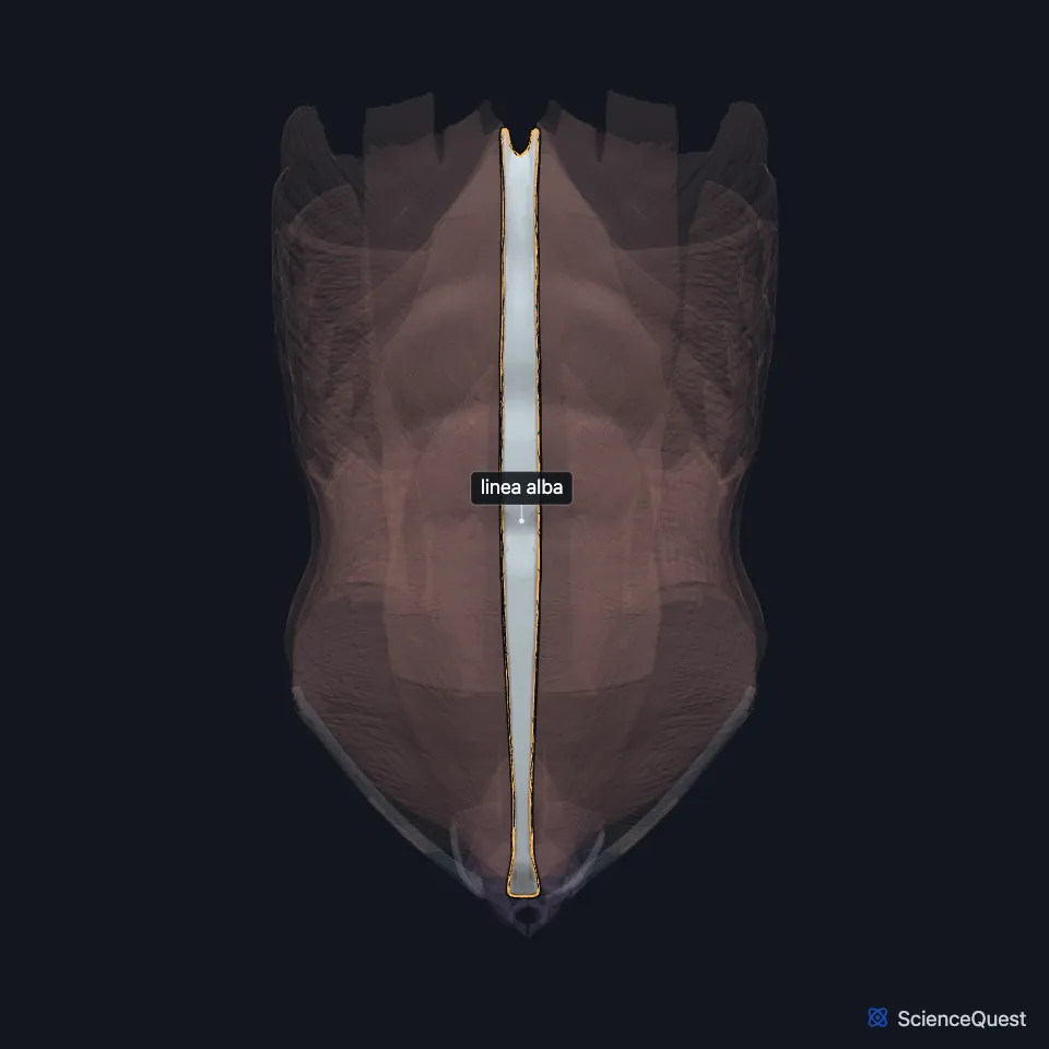
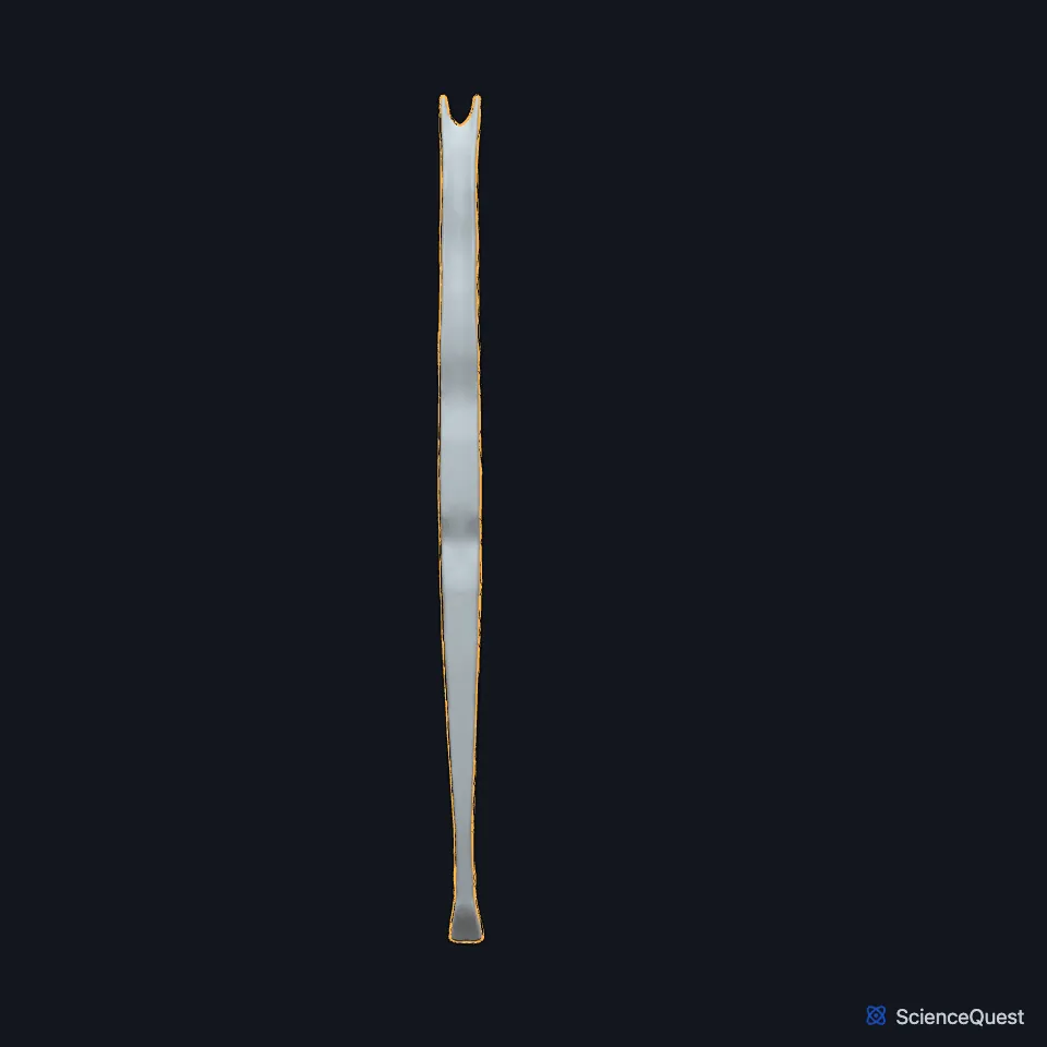
Open it in the Abdominal Muscles Explorer See it alone in 3D
Where is the linea alba?
It lies between the inner borders of the two rectus abdominis muscles and separates one from the other. (Gray 1918, 6d. Muscles of the Abdomen)
At its lower end, its surface fibers pass in front of the rectus heads to the symphysis. Its deep fibers form a triangular sheet behind them, the adminiculum lineae albae, attached to the pubic crest. (Gray 1918, 6d. Muscles of the Abdomen)
The umbilicus lies in it. In the fetus the umbilical opening carries the umbilical vessels, and in the adult it is closed. (Gray 1918, 6d. Muscles of the Abdomen)
What is the linea alba made of?
The aponeuroses of the external oblique, the internal oblique and the transversus abdominis blend in the midline to form it. (StatPearls, Abdominal Wall; Gray 1918, 6d. Muscles of the Abdomen)
Most studies in a 2026 systematic review describe layered collagen, with oblique fibers in front and transverse fibers behind. (Martonovich et al. 2026, Plast Reconstr Surg Glob Open)
What tenses the linea alba, and what part does it play in surgery and hernias?
The pyramidalis tenses it from below. (StatPearls, Abdominal Wall)
Surgeons open the abdomen through it in a midline incision. It carries few blood vessels, so the cut bleeds little and divides no nerves. (StatPearls, Anterolateral Abdominal Wall Muscles)
Epigastric hernias push through it above the umbilicus. In diastasis recti it thins and widens and bulges when pressure in the abdomen rises, though it is no true hernia. (StatPearls, Abdominal Wall and Anterolateral Abdominal Wall Fascia)
What passes through or joins the linea alba?
Small openings in it let vessels and nerves through. (Gray 1918, 6d. Muscles of the Abdomen)
Fibers of the reflected inguinal ligament climb from each side and cross at the linea alba, in front of the pyramidalis. (StatPearls, Inguinal Ligament; Gray 1918, 6d. Muscles of the Abdomen)
Where the sources differ
Gray’s 1918 text has the linea alba narrow below and broader above, where the recti diverge. A 2026 systematic review found it widest at the umbilicus, intermediate above it and narrowest near the pubic symphysis. (Gray 1918, 6d. Muscles of the Abdomen; Martonovich et al. 2026, Plast Reconstr Surg Glob Open)
Gray’s 1918 text has each external oblique aponeurosis interlacing with its partner at the midline. The 2026 review found that the two newer studies to test this saw no fibers cross the midline, and it set aside older reports of an X-shaped crossing for high risk of bias. (Gray 1918, 6d. Muscles of the Abdomen; Martonovich et al. 2026, Plast Reconstr Surg Glob Open)
| Feature | Detail and source |
|---|---|
| Extent | Xiphoid process to the pubic symphysis (Gray 1918, 6d. Muscles of the Abdomen) |
| Formed by | Aponeuroses of the external oblique, internal oblique and transversus abdominis of both sides (StatPearls, Abdominal Wall; Gray 1918, 6d. Muscles of the Abdomen) |
| Lies between | The two rectus abdominis muscles (Gray 1918, 6d. Muscles of the Abdomen) |
| Widest | At the umbilicus, in a 2026 systematic review (Martonovich et al. 2026, Plast Reconstr Surg Glob Open) |
Touches in this model
Measured on the BodyParts3D meshes: surfaces within 2 mm of the linea alba, nearest first, 12 of 22.
- Left external oblique in contact
- Left internal oblique in contact
- Left pyramidalis in contact
- Left rectus abdominis in contact
- Left transversus abdominis in contact
- Right external oblique in contact
- Right internal oblique in contact
- Right pyramidalis in contact
- Right rectus abdominis in contact
- Right transversus abdominis in contact
- Jejunum in contact
- Left hip bone in contact
Sources
Sources read
- Gray’s Anatomy of the Human Body, 20th US edition (1918), IV. Myology, 6d. The Muscles and Fasciae of the Abdomen. Henry Gray. Public domain. Read .
- StatPearls, Anatomy, Abdomen and Pelvis: Abdominal Wall. William Flynn; Paula Vickerton. Last update . CC BY-NC-ND 4.0. Read .
- Plastic and Reconstructive Surgery Global Open, The Anatomy of the Human Linea Alba: A Systematic Review and Meta-analysis, Plast Reconstr Surg Glob Open 14, e7935 (full text, PMC13441126, doi 10.1097/GOX.0000000000007935). Martonovich N, Fede C, De Bon G, Sun Y, De Luca C, Stecco C, Behrbalk E. CC BY-NC-ND 4.0. Read . Rechecked .
- StatPearls, Anatomy, Anterolateral Abdominal Wall Muscles. Matthew A. Varacallo; Stephanie Scharbach; Mohammed A. Al-Dhahir. Last update . CC BY-NC-ND 4.0. Read .
- StatPearls, Anatomy, Abdomen and Pelvis: Anterolateral Abdominal Wall Fascia. Leandra A. Jelinek; Stephanie Scharbach; Sarang Kashyap; Troy Ferguson. Last update . CC BY-NC-ND 4.0. Read .
- StatPearls, Anatomy, Abdomen and Pelvis: Inguinal Ligament (Crural Ligament. Poupart Ligament). Kavin Sugumar; Mohit Gupta. Last update . CC BY-NC-ND 4.0. Read .
The values are facts and free to use; this page’s selection and presentation are © 2026 ScienceQuest.