Inguinal ligament
The inguinal ligament is the thick, in-folded lower border of the aponeurosis of the external oblique. It spans from the anterior superior iliac spine to the pubic tubercle. (Gray 1918, 6d. Muscles of the Abdomen; StatPearls, Inguinal Ligament)
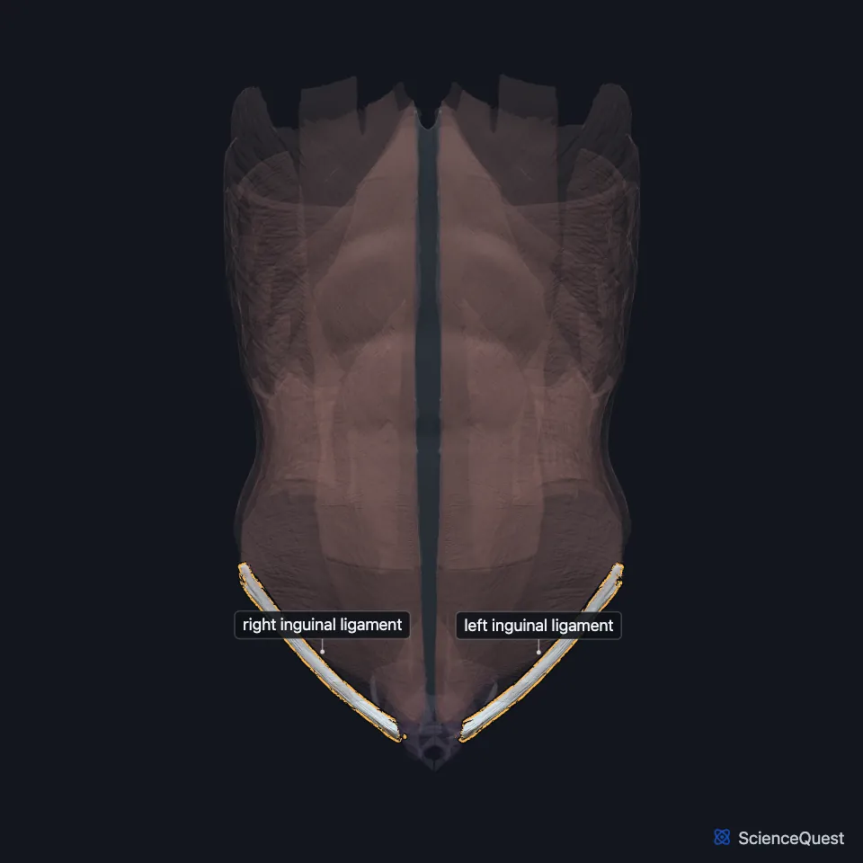
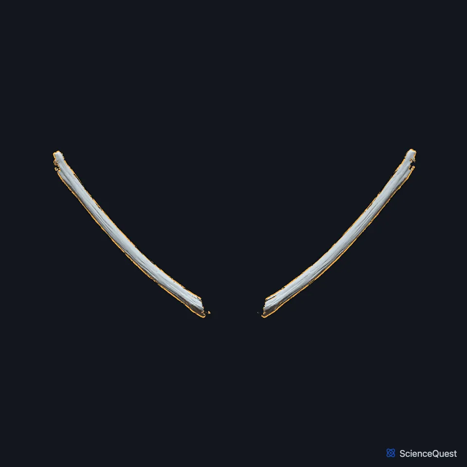
Open it in the Abdominal Muscles Explorer See it alone in 3D
Where is the inguinal ligament?
It joins the fascia lata of the thigh below, which gives it a downward curve. Its outer part is rounded and oblique, and its inner part is grooved and lies more horizontal. (StatPearls, Inguinal Ligament; Gray 1918, 6d. Muscles of the Abdomen)
Its grooved upper surface forms the floor of the inguinal canal. The canal carries the ilioinguinal nerve with the spermatic cord in males and the round ligament in females. (StatPearls, Inguinal Ligament; Gray 1918, 6d. Muscles of the Abdomen)
How is the inguinal ligament built?
At its inner end it fans out into the lacunar ligament, whose apex sits at the pubic tubercle and whose back edge attaches to the pecten pubis. The pectineal ligament runs outward from the base of the lacunar ligament. (StatPearls, Inguinal Ligament)
Anatomists still debate whether it counts as a ligament or a tendon. It arises from a muscle’s aponeurosis, and it stabilizes no joint. (StatPearls, Inguinal Ligament)
Falloppio first described it in 1562, and the French anatomist François Poupart described it further. Gray’s 1918 text calls it Poupart’s ligament, and the Foundational Model of Anatomy also lists Fallopian ligament. (StatPearls, Inguinal Ligament; Gray 1918, 6d. Muscles of the Abdomen; FMA, Inguinal ligament)
What does the inguinal ligament do?
It anchors muscles: the internal oblique arises from its outer two thirds, the transversus abdominis from its outer third and the cremaster from its middle third. (StatPearls, Inguinal Ligament)
Clinicians use it to tell groin hernias apart. A bulge above it is an inguinal hernia, and one below it is a femoral hernia. (StatPearls, Inguinal Ligament)
What passes under the inguinal ligament?
It bridges the structures passing from the pelvis to the thigh. From lateral to medial, the lateral femoral cutaneous nerve, the femoral nerve, the external iliac artery, the femoral vein and the femoral ring pass deep to it. The iliopsoas, formed by the iliacus and the psoas major, fills the space bounded by the ligament, the iliopectineal band and the front border of the iliac bone. (StatPearls, Inguinal Ligament and Psoas Major)
With the sartorius and adductor longus, it bounds the femoral triangle. The deep inguinal ring lies half an inch above its midpoint. (StatPearls, Inguinal Ligament)
Does the inguinal ligament vary?
A 2009 cadaver study that StatPearls cites found a reflected inguinal ligament in about 83% of bodies. (StatPearls, Inguinal Ligament)
Where the sources differ
StatPearls has the internal oblique arising from the outer two thirds of the ligament. Gray’s 1918 text gives the outer half of its grooved upper surface. (StatPearls, Inguinal Ligament; Gray 1918, 6d. Muscles of the Abdomen)
| Feature | Detail and source |
|---|---|
| Attachments | Anterior superior iliac spine to the pubic tubercle (StatPearls, Inguinal Ligament; Gray 1918, 6d. Muscles of the Abdomen) |
| Formed by | Lower border of the external oblique aponeurosis (Gray 1918, 6d. Muscles of the Abdomen) |
| Continuous with | Fascia lata of the thigh, below (StatPearls, Inguinal Ligament) |
| Gives origin to | Internal oblique, transversus abdominis and cremaster (StatPearls, Inguinal Ligament) |
| Other names | Poupart’s ligament, Fallopian ligament (Gray 1918, 6d. Muscles of the Abdomen; FMA, Inguinal ligament) |
Touches in this model
Measured on the BodyParts3D meshes: surfaces within 2 mm of the inguinal ligament, nearest first, 12 of 25.
- Left external oblique in contact
- Left internal oblique in contact
- Left pyramidalis in contact
- Left transversus abdominis in contact
- Right external oblique in contact
- Right internal oblique in contact
- Right pyramidalis in contact
- Right transversus abdominis in contact
- Left adductor longus in contact
- Left deferent duct in contact
- Left external iliac artery in contact
- Left external iliac vein in contact
Sources
Sources read
- Gray’s Anatomy of the Human Body, 20th US edition (1918), IV. Myology, 6d. The Muscles and Fasciae of the Abdomen. Henry Gray. Public domain. Read .
- StatPearls, Anatomy, Abdomen and Pelvis: Inguinal Ligament (Crural Ligament. Poupart Ligament). Kavin Sugumar; Mohit Gupta. Last update . CC BY-NC-ND 4.0. Read .
- Foundational Model of Anatomy, FMA19855, Inguinal ligament (read on EBI OLS). FMA release terms, University of Washington. Read .
- StatPearls, Anatomy, Bony Pelvis and Lower Limb: Psoas Major. Marco A. Siccardi; Muhammad Ali Tariq; Cristina Valle. Last update . CC BY-NC-ND 4.0. Read .
The values are facts and free to use; this page’s selection and presentation are © 2026 ScienceQuest.