External iliac artery
The external iliac artery is the larger of the two branches of the common iliac artery and the main supply route to the lower limb. Below the inguinal ligament it becomes the femoral artery. (StatPearls, Abdomen and Pelvis: External Iliac Arteries; Gray’s Anatomy (1918), The External Iliac Artery)
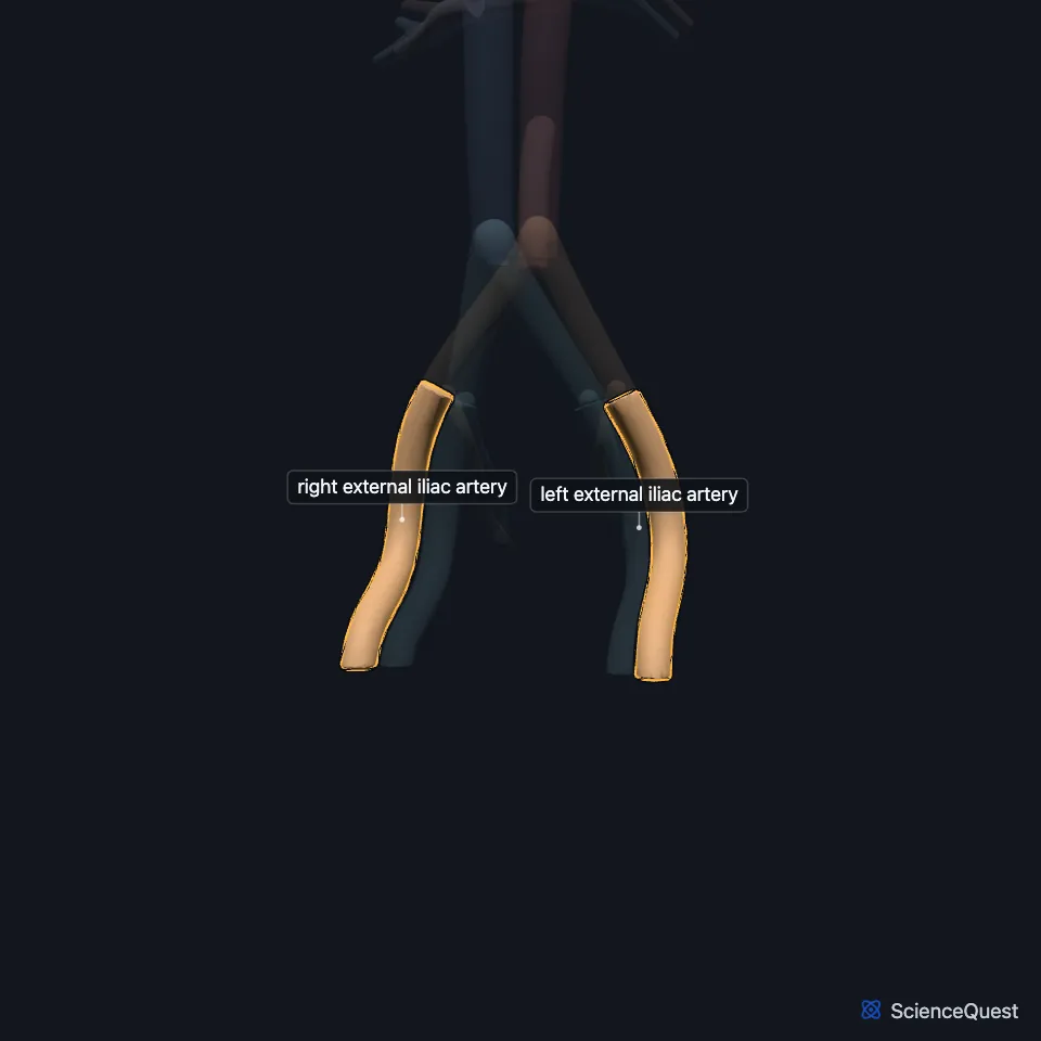
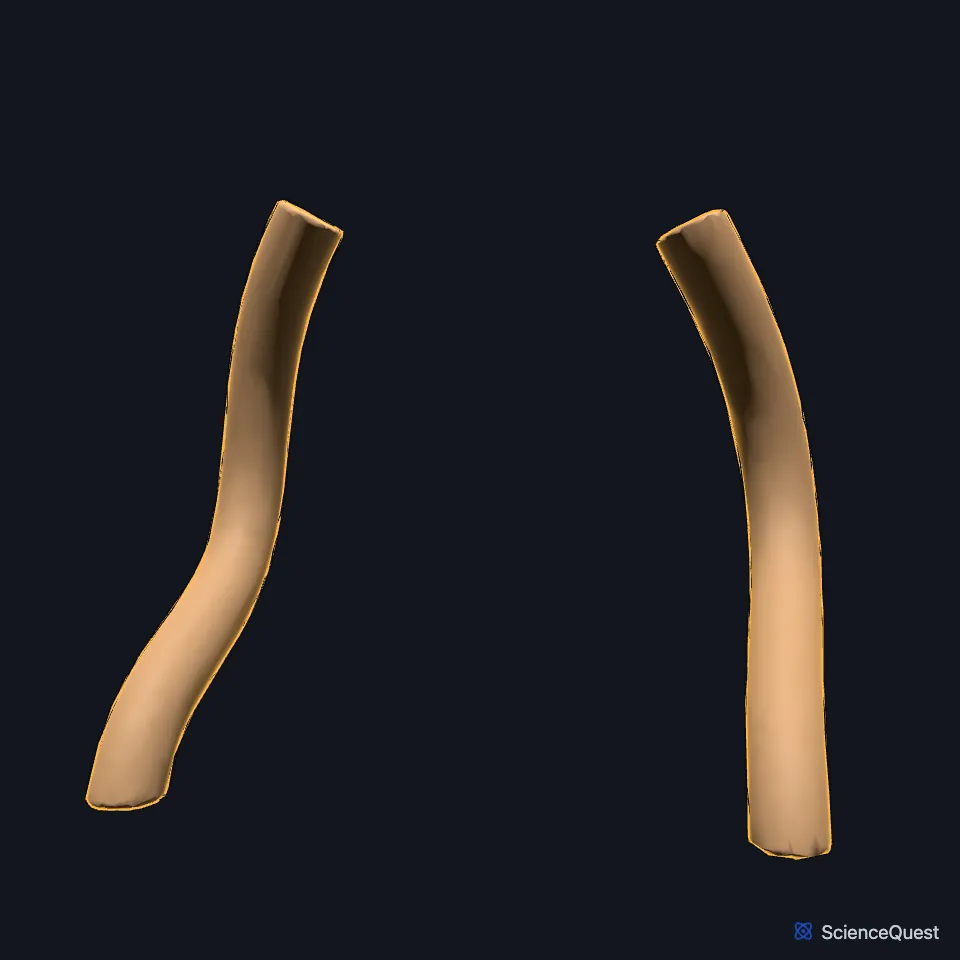
Where is the external iliac artery?
Each begins where the common iliac artery divides, in front of the sacroiliac joint, and runs along the medial border of the psoas major to leave the pelvis behind the inguinal ligament. (StatPearls, Abdomen and Pelvis: External Iliac Arteries)
What does the external iliac artery do?
It is mostly a short conduit between the common iliac and common femoral arteries. StatPearls has it supply no muscle or tissue directly, leaving that to its two branches. (StatPearls, Abdomen and Pelvis: External Iliac Arteries)
The inferior epigastric artery leaves its medial side and climbs behind the rectus abdominis. The deep circumflex iliac artery leaves its lateral side and runs along the iliac crest. (StatPearls, Abdomen and Pelvis: External Iliac Arteries; Gray’s Anatomy (1918), The External Iliac Artery)
The inferior epigastric artery supplies the rectus abdominis, and the deep circumflex iliac artery the transversus abdominis and internal oblique. (StatPearls, Abdomen and Pelvis: External Iliac Arteries)
What lies next to the external iliac artery?
The external iliac vein lies wholly on the medial side of the artery on the left. On the right, Gray’s Anatomy (1918) has the vein start medial to the artery and drift behind it as it rises. (Gray’s Anatomy (1918), The Veins of the Abdomen and Pelvis)
At its origin the ovarian vessels cross it in women, and sometimes the ureter. Near its end the ductus deferens in men and the round ligament of the uterus in women curve across its medial side. (Gray’s Anatomy (1918), The External Iliac Artery)
Eight to ten external iliac lymph nodes lie around it, in lateral, medial and anterior groups. (StatPearls, Abdomen and Pelvis: External Iliac Arteries)
Why does the external iliac artery matter in vascular surgery?
Wires and sheaths for endovascular repair of an abdominal aortic aneurysm usually pass from the common femoral artery through the external and common iliac arteries. A very narrow, twisted or calcified external iliac artery may force another route or open surgery. (StatPearls, Abdomen and Pelvis: External Iliac Arteries)
The corona mortis, a link between the obturator vessels and the external iliac or inferior epigastric vessels, is found in 9.8% of patients. If it is cut in pelvic surgery, the vessel can retract and be hard to control. (StatPearls, Abdomen and Pelvis: External Iliac Arteries)
Where the sources differ
The sources place its exit under the inguinal ligament differently. StatPearls puts it about one-third of the way from the pubic tubercle to the anterior superior iliac spine; Gray’s Anatomy (1918) puts it midway between the anterior superior iliac spine and the pubic symphysis. (StatPearls, Abdomen and Pelvis: External Iliac Arteries; Gray’s Anatomy (1918), The External Iliac Artery)
The sources differ on whether it feeds any tissue itself. StatPearls says it supplies no muscle or tissue directly; Gray’s Anatomy (1918) gives it several small branches to the psoas major and the nearby lymph glands (lymph nodes), besides its two large ones. (StatPearls, Abdomen and Pelvis: External Iliac Arteries; Gray’s Anatomy (1918), The External Iliac Artery)
| Feature | Detail and source |
|---|---|
| Arises from | Common iliac artery (StatPearls, Abdomen and Pelvis: External Iliac Arteries) |
| Branches | Inferior epigastric and deep circumflex iliac arteries (StatPearls, Abdomen and Pelvis: External Iliac Arteries) |
| Continues as | Common femoral artery, below the inguinal ligament (StatPearls, Abdomen and Pelvis: External Iliac Arteries; Gray’s Anatomy (1918), The External Iliac Artery) |
Touches in this model
Measured on the BodyParts3D meshes: surfaces within 2 mm of the external iliac artery, nearest first, 12 of 23.
- Left common iliac artery in contact
- Left internal iliac artery in contact
- Right common iliac artery in contact
- Right internal iliac artery in contact
- Ileum in contact
- Left deferent duct in contact
- Left external oblique in contact
- Left inguinal ligament in contact
- Left psoas major in contact
- Left transversus abdominis in contact
- Right deferent duct in contact
- Right external oblique in contact
Sources
Sources read
- StatPearls, Anatomy, Abdomen and Pelvis: External Iliac Arteries. Eric Hammond; Ali Nassereddin; Michael Costanza. Last update . CC BY-NC-ND 4.0. Read .
- Anatomy of the Human Body (Gray’s Anatomy), 20th US edition, 1918, Angiology, The Arteries of the Lower Extremity, The External Iliac Artery (A. Iliaca Externa), page 622; Internet Archive scan of the 1918 Lea and Febiger printing, terminology of 1918 (internal spermatic vessels for testicular vessels, hypogastric for internal iliac). Henry Gray; edited by Warren H. Lewis. Public domain. Read .
- Anatomy of the Human Body (Gray’s Anatomy), 20th US edition, 1918, Angiology, The Veins of the Lower Extremity, Abdomen, and Pelvis: the external iliac, hypogastric and common iliac veins, and the renal veins among the tributaries of the inferior vena cava, pages 672 to 679; Internet Archive scan of the 1918 Lea and Febiger printing, terminology of 1918 (hypogastric for internal iliac, spermatic for testicular, hemorrhoidal for rectal). Henry Gray; edited by Warren H. Lewis. Public domain. Read .
The values are facts and free to use; this page’s selection and presentation are © 2026 ScienceQuest.