Common iliac vein
Each common iliac vein forms from the external and internal iliac veins in front of the sacroiliac joint. The right and left veins join at the fifth lumbar vertebra to form the inferior vena cava. (Gray’s Anatomy (1918), The Veins of the Abdomen and Pelvis; StatPearls, Abdomen and Pelvis: Inferior Vena Cava)
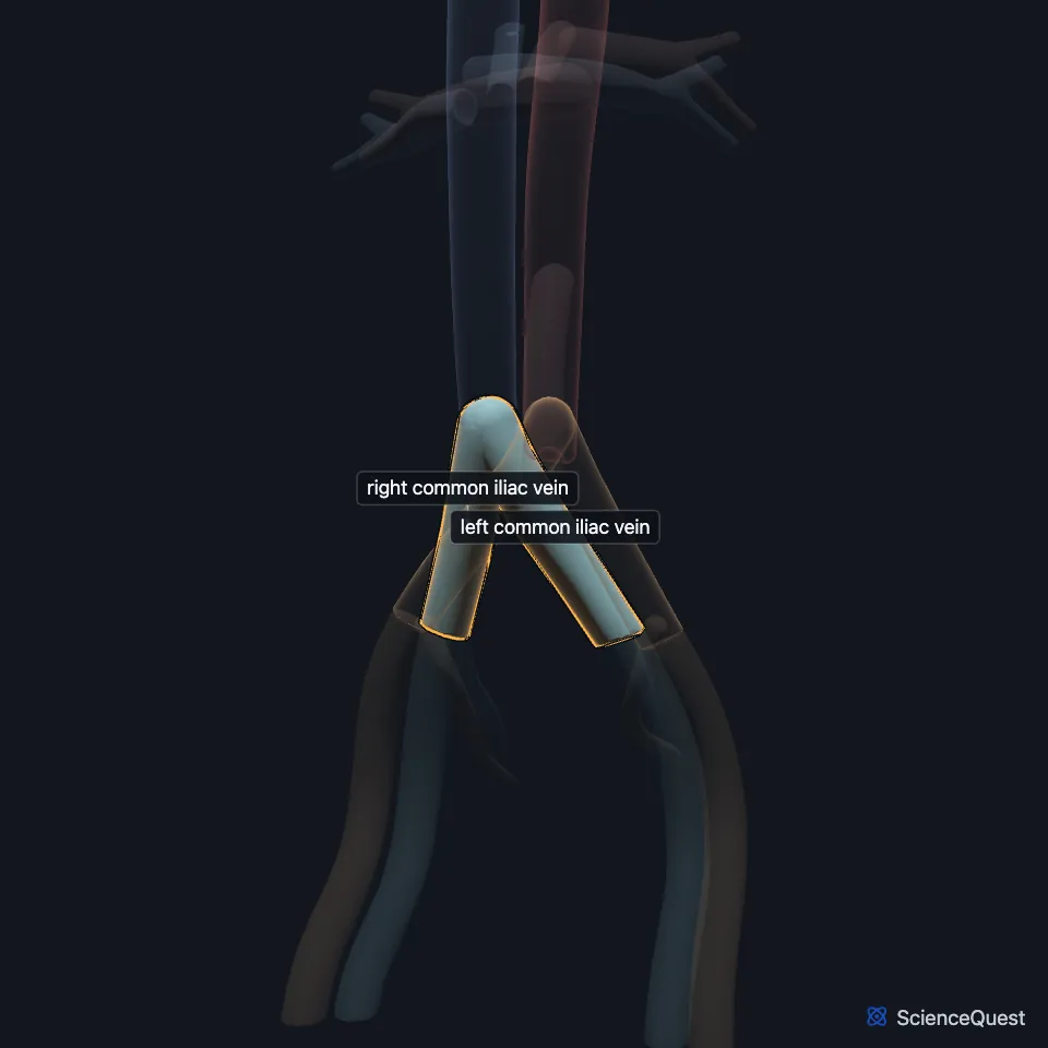
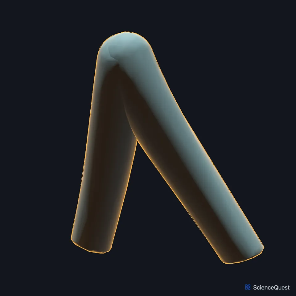
Where are the common iliac veins?
The right vein is shorter and almost vertical, and rises behind and then lateral to the right common iliac artery. (Gray’s Anatomy (1918), The Veins of the Abdomen and Pelvis)
The left vein is longer and more oblique. It lies first on the medial side of its own artery and then passes behind the right common iliac artery. (Gray’s Anatomy (1918), The Veins of the Abdomen and Pelvis)
What do the common iliac veins drain?
They carry blood from the lower limbs, which reaches them from the femoral and external iliac veins, and from the pelvis, through the internal iliac veins, into the inferior vena cava. (StatPearls, Abdomen and Pelvis: Inferior Vena Cava and Abdomen and Pelvis, Pelvis)
Each receives the iliolumbar vein and sometimes lateral sacral veins, and the left also receives the middle sacral vein. Gray’s Anatomy (1918) found no valves in them. (Gray’s Anatomy (1918), The Veins of the Abdomen and Pelvis)
What lies next to the common iliac veins?
Because the left vein passes behind the right common iliac artery, that artery can press it against the lower lumbar spine. (Gray’s Anatomy (1918), The Veins of the Abdomen and Pelvis; StatPearls, May-Thurner Syndrome)
The superior rectal branch of the inferior mesenteric artery crosses in front of the left common iliac artery and vein as it enters the pelvis. (StatPearls, Abdomen and Pelvis: Inferior Mesenteric Artery)
What is May-Thurner syndrome?
May-Thurner syndrome is a symptomatic blockage of leg venous return from compression of a common iliac vein, most often the left vein by the right common iliac artery. The artery’s pulsation damages the vein lining and builds fibrous bands or spurs that narrow it and can lead to deep vein thrombosis. (StatPearls, May-Thurner Syndrome)
It should be considered in anyone with a deep vein thrombosis of the iliac and femoral veins. CT venography detects iliac vein compression with more than 95% sensitivity and specificity. (StatPearls, May-Thurner Syndrome)
How does the left common iliac vein develop?
In the embryo, the sacrocardinal veins join to form the left common iliac vein. (StatPearls, Abdomen and Pelvis: Inferior Vena Cava)
| Feature | Detail and source |
|---|---|
| Formed by | External iliac vein and internal iliac vein (StatPearls, Abdomen and Pelvis, Pelvis) |
| End in | Inferior vena cava, at about the fifth lumbar vertebra (StatPearls, Abdomen and Pelvis: Inferior Vena Cava) |
| Valves | None found (Gray’s Anatomy, 1918) (Gray’s Anatomy (1918), The Veins of the Abdomen and Pelvis) |
Touches in this model
Measured on the BodyParts3D meshes: surfaces within 2 mm of the common iliac vein, nearest first, 12 of 14.
- Inferior vena cava in contact
- Left external iliac vein in contact
- Left internal iliac vein in contact
- Right common iliac artery in contact
- Right external iliac vein in contact
- Right internal iliac vein in contact
- Fifth lumbar vertebra in contact
- Fourth lumbar vertebra in contact
- Intervertebral disk of fifth lumbar vertebra in contact
- Intervertebral disk of fourth lumbar vertebra in contact
- Right psoas major in contact
- Skin in contact
Sources
Sources read
- Anatomy of the Human Body (Gray’s Anatomy), 20th US edition, 1918, Angiology, The Veins of the Lower Extremity, Abdomen, and Pelvis: the external iliac, hypogastric and common iliac veins, and the renal veins among the tributaries of the inferior vena cava, pages 672 to 679; Internet Archive scan of the 1918 Lea and Febiger printing, terminology of 1918 (hypogastric for internal iliac, spermatic for testicular, hemorrhoidal for rectal). Henry Gray; edited by Warren H. Lewis. Public domain. Read .
- StatPearls, Anatomy, Abdomen and Pelvis: Inferior Vena Cava. William D. Tucker; Ribesh Shrestha; Bracken Burns. Last update . CC BY-NC-ND 4.0. Read .
- StatPearls, Anatomy, Abdomen and Pelvis, Pelvis. Shazia R. Chaudhry; Ahmed Nahian; Khalid Chaudhry. Last update . CC BY-NC-ND 4.0. Read .
- StatPearls, May-Thurner Syndrome. Ankit Mangla; Hussein Hamad. Last update . CC BY-NC-ND 4.0. Read .
- StatPearls, Anatomy, Abdomen and Pelvis: Inferior Mesenteric Artery. Adekunle E. Omole; Kelly Van Fossen. Last update . CC BY-NC-ND 4.0. Read .
The values are facts and free to use; this page’s selection and presentation are © 2026 ScienceQuest.