Inferior mesenteric artery
The inferior mesenteric artery is the artery of the hindgut. It leaves the abdominal aorta at about the third lumbar vertebra and has three main branches: the left colic, sigmoid and superior rectal arteries. (StatPearls, Abdomen and Pelvis: Inferior Mesenteric Artery)
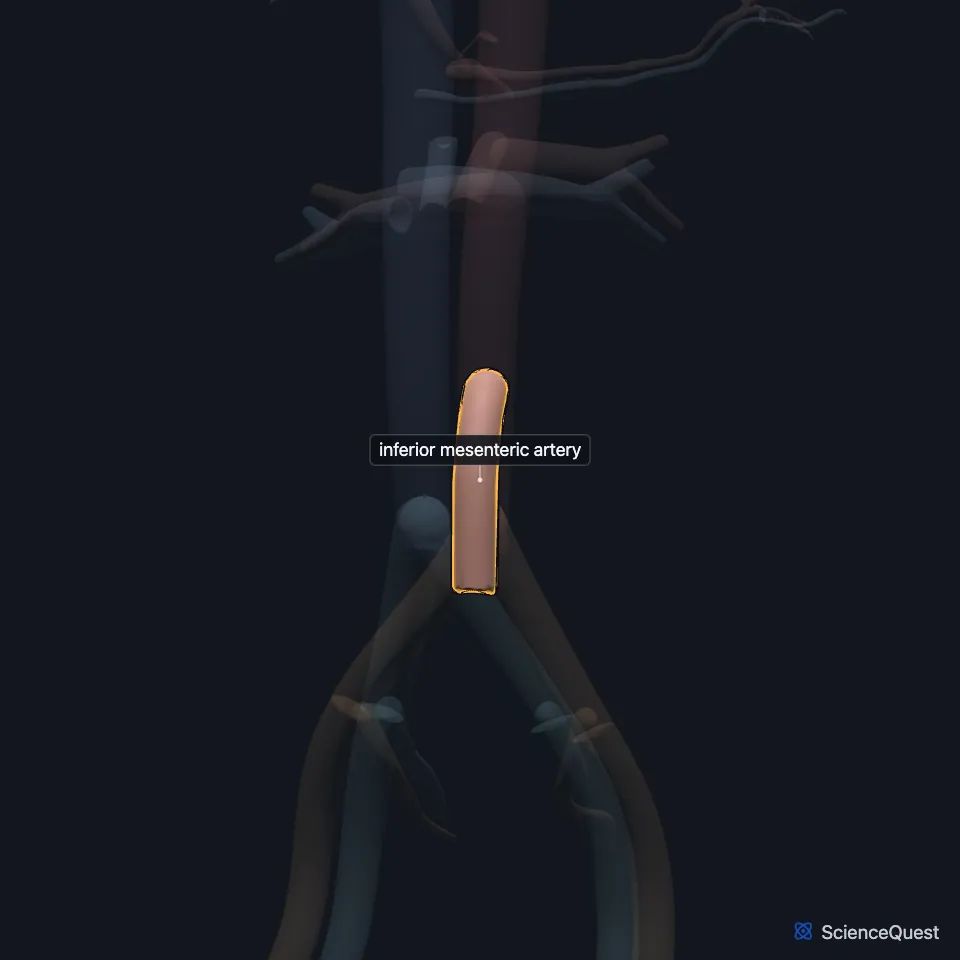
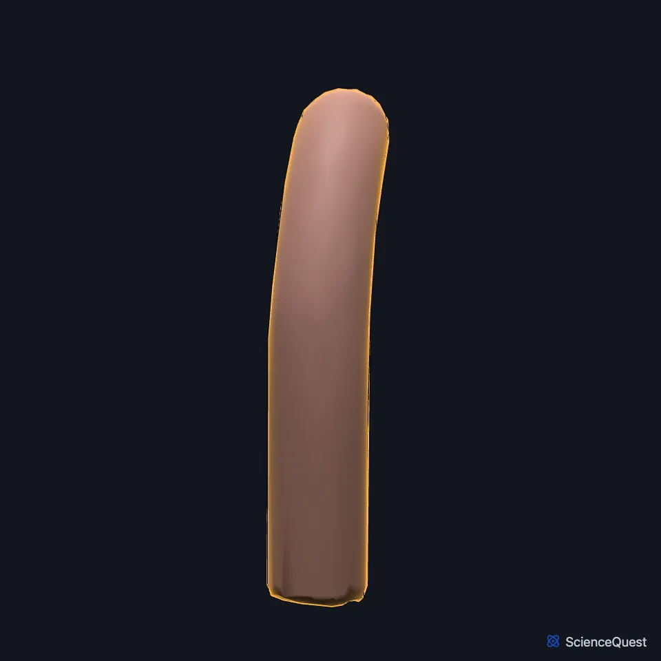
Where is the inferior mesenteric artery?
It arises from the front and left side of the aorta, below the renal arteries and above the aortic bifurcation, and runs behind the third part of the duodenum. (StatPearls, Abdomen and Pelvis: Inferior Mesenteric Artery)
Gray’s Anatomy (1918) places its origin about 3 or 4 cm above the aortic bifurcation. It descends behind the peritoneum, first in front of and then to the left of the aorta, and crosses the left common iliac artery to enter the pelvis as the superior hemorrhoidal artery, the 1918 name for the superior rectal artery. (Gray’s Anatomy (1918), The Abdominal Aorta)
What does the inferior mesenteric artery supply?
It supplies the hindgut: the distal third of the transverse colon, the descending and sigmoid colon and the upper rectum. (StatPearls, Abdomen and Pelvis: Inferior Mesenteric Artery)
Each branch reaches the colon through arcades of small arteries. The superior rectal artery, its last branch, begins at the pelvic brim, supplies the upper rectum and links with the middle and inferior rectal arteries. (StatPearls, Abdomen and Pelvis: Inferior Mesenteric Artery)
What lies next to the inferior mesenteric artery?
The inferior mesenteric vein runs with it but usually drains into the splenic vein, not the inferior vena cava. Gray’s Anatomy (1918) adds that it sometimes ends where the splenic and superior mesenteric veins join. (StatPearls, Abdomen and Pelvis: Inferior Mesenteric Artery; Gray’s Anatomy (1918), The Portal System of Veins)
Its left colic branch passes in front of the left ureter and the psoas major, and the superior rectal artery passes in front of the left common iliac artery and vein. (StatPearls, Abdomen and Pelvis: Inferior Mesenteric Artery)
A horseshoe kidney stays low in the abdomen because the fused kidneys, rising in the embryo, are caught below the inferior mesenteric artery. (StatPearls, Abdomen and Pelvis: Inferior Mesenteric Artery)
Where does the inferior mesenteric artery meet the superior mesenteric artery?
The marginal artery of Drummond links the inferior and superior mesenteric systems along the colon. Fewer than half of people have an unbroken channel around the splenic flexure. (StatPearls, Abdomen and Pelvis: Inferior Mesenteric Artery)
Watershed areas get blood from the farthest branches of two or more arteries and are especially vulnerable to ischemia in shock or heart failure. The splenic flexure, fed by both mesenteric arteries, and the rectosigmoid junction, fed by this artery and the internal iliac arteries, are two of them. (StatPearls, Abdomen and Pelvis: Inferior Mesenteric Artery)
How does the inferior mesenteric artery develop?
The celiac trunk and the superior and inferior mesenteric arteries persist from the 10th, 13th and 21st ventral segmental arteries of the embryo. (StatPearls, Abdomen and Pelvis: Celiac Trunk)
Where the sources differ
The sources draw its territory differently. StatPearls gives the distal third of the transverse colon and the proximal two-thirds of the rectum; Gray’s Anatomy (1918) gives the left half of the transverse colon and the greater part of the rectum. (StatPearls, Abdomen and Pelvis: Inferior Mesenteric Artery; Gray’s Anatomy (1918), The Abdominal Aorta)
| Feature | Detail and source |
|---|---|
| Arises from | Abdominal aorta, at about the level of the third lumbar vertebra (L3) (StatPearls, Abdomen and Pelvis: Inferior Mesenteric Artery) |
| Branches | Left colic, sigmoid and superior rectal arteries (StatPearls, Abdomen and Pelvis: Inferior Mesenteric Artery) |
| Vein | Inferior mesenteric vein, which usually drains into the splenic vein (StatPearls, Abdomen and Pelvis: Inferior Mesenteric Artery; Gray’s Anatomy (1918), The Portal System of Veins) |
Touches in this model
Measured on the BodyParts3D meshes: surfaces within 2 mm of the inferior mesenteric artery, nearest first.
- Descending aorta in contact
Sources
Sources read
- StatPearls, Anatomy, Abdomen and Pelvis: Inferior Mesenteric Artery. Adekunle E. Omole; Kelly Van Fossen. Last update . CC BY-NC-ND 4.0. Read .
- Anatomy of the Human Body (Gray’s Anatomy), 20th US edition, 1918, Angiology, The Abdominal Aorta, its branches and the middle sacral artery; Internet Archive scan of the 1918 Lea and Febiger printing, terminology of 1918. Henry Gray; edited by Warren H. Lewis. Public domain. Read .
- Anatomy of the Human Body (Gray’s Anatomy), 20th US edition, 1918, Angiology, The Portal System of Veins, the portal vein; Internet Archive scan of the 1918 Lea and Febiger printing, terminology of 1918. Henry Gray; edited by Warren H. Lewis. Public domain. Read .
- StatPearls, Anatomy, Abdomen and Pelvis: Celiac Trunk. Dr. Rohin Garg; Ali Nassereddin; Tafline C. Arbor. Last update . CC BY-NC-ND 4.0. Read .
The values are facts and free to use; this page’s selection and presentation are © 2026 ScienceQuest.