Splenic vein
The splenic vein drains the spleen. It runs behind the pancreas and joins the superior mesenteric vein to form the portal vein. (StatPearls, Spleen; Gray’s Anatomy (1918), The Portal System of Veins)
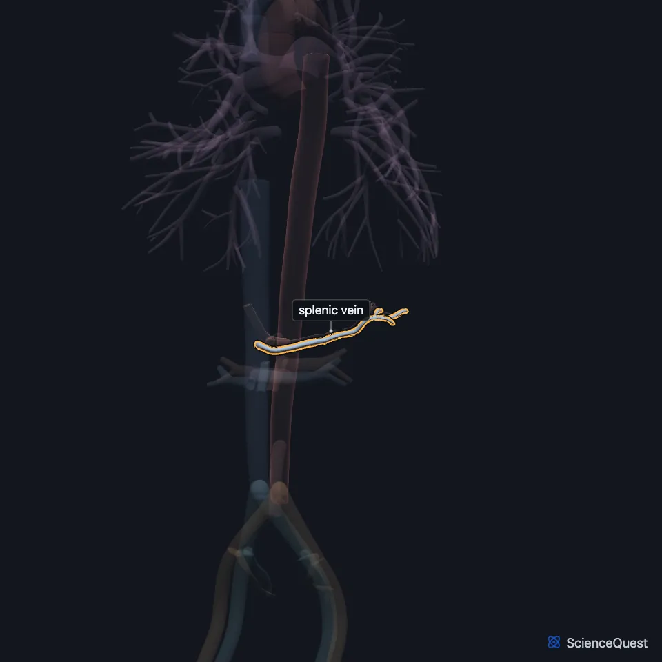
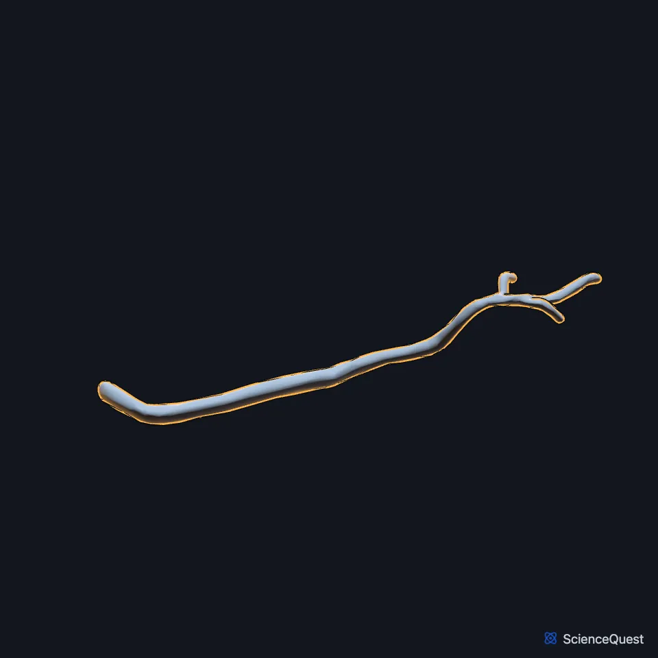
Where is the splenic vein?
It starts as five or six large branches leaving the spleen. The single vein they form runs from left to right in a groove on the upper back part of the pancreas, below the splenic artery. (Gray’s Anatomy (1918), The Portal System of Veins)
It ends behind the neck of the pancreas, where it meets the superior mesenteric vein at a right angle. (Gray’s Anatomy (1918), The Portal System of Veins; StatPearls, Liver)
What does the splenic vein drain?
It carries blood from the spleen and from the body and neck of the pancreas into the portal vein, which takes it to the liver. (StatPearls, Pancreas and Abdomen and Pelvis: Hepatoduodenal Ligament)
Its tributaries are the short gastric and left gastroepiploic veins from the stomach, the pancreatic veins and the inferior mesenteric vein. (Gray’s Anatomy (1918), The Portal System of Veins; StatPearls, Stomach and Abdomen and Pelvis: Inferior Mesenteric Artery)
Gray’s Anatomy (1918) adds that the inferior mesenteric vein sometimes ends instead in the angle where the splenic and superior mesenteric veins meet. (Gray’s Anatomy (1918), The Portal System of Veins)
The portal vein it helps form brings about 75% of the liver’s blood; the hepatic artery brings the rest. (StatPearls, Abdomen and Pelvis, Portal Venous System)
What lies next to the splenic vein?
It lies behind the pancreas and below the splenic artery, and the two vessels pass through the hilum of the spleen. (StatPearls, Spleen; Gray’s Anatomy (1918), The Abdominal Aorta)
Gray’s Anatomy (1918), which calls it the lienal vein, notes that it is large but runs straight, unlike the winding artery. (Gray’s Anatomy (1918), The Portal System of Veins)
What happens to the splenic vein in portal hypertension?
As portal hypertension worsens, congestion of the splenic vein can enlarge the spleen. A splenorenal shunt can sometimes ease portal hypertension by reducing the blood the splenic vein contributes. (StatPearls, Physiology, Spleen)
| Feature | Detail and source |
|---|---|
| Begins | Hilum of the spleen (StatPearls, Spleen) |
| Ends | Joins the superior mesenteric vein behind the neck of the pancreas to form the portal vein (StatPearls, Liver) |
| Tributaries | Short gastric, left gastroepiploic, pancreatic and inferior mesenteric veins (Gray’s Anatomy (1918), The Portal System of Veins) |
Touches in this model
Measured on the BodyParts3D meshes: surfaces within 2 mm of the splenic vein, nearest first.
- Inferior vena cava in contact
- Splenic artery in contact
- Pancreas in contact
- Spleen in contact
- Celiac artery 0.4 mm away
Sources
Sources read
- StatPearls, Anatomy, Abdomen and Pelvis, Spleen. Adekunle E. Omole; Marjorie V. Launico. Last update . CC BY-NC-ND 4.0. Read .
- Anatomy of the Human Body (Gray’s Anatomy), 20th US edition, 1918, Angiology, The Portal System of Veins, the portal vein; Internet Archive scan of the 1918 Lea and Febiger printing, terminology of 1918. Henry Gray; edited by Warren H. Lewis. Public domain. Read .
- StatPearls, Anatomy, Abdomen and Pelvis: Liver. Hamilton Vernon; Chase J. Wehrle; Valentine Sampson K. Alia; Anup Kasi. Last update . CC BY-NC-ND 4.0. Read .
- StatPearls, Anatomy, Abdomen and Pelvis, Pancreas. Saurabh S. Talathi; Adekunle E. Omole; Michael Young. Last update . CC BY-NC-ND 4.0. Read .
- StatPearls, Anatomy, Abdomen and Pelvis: Hepatoduodenal Ligament. Justyn Nakashima; Bruno Bordoni. Last update . CC BY-NC-ND 4.0. Read .
- StatPearls, Anatomy, Abdomen and Pelvis: Stomach. Shazia R. Chaudhry; Maria Nataly P. Liman; Adekunle E. Omole; Diana C. Peterson. Last update . CC BY-NC-ND 4.0. Read .
- StatPearls, Anatomy, Abdomen and Pelvis: Inferior Mesenteric Artery. Adekunle E. Omole; Kelly Van Fossen. Last update . CC BY-NC-ND 4.0. Read .
- StatPearls, Anatomy, Abdomen and Pelvis, Portal Venous System (Hepatic Portal System). Joshua M. Harkins; Bareeqah Ahmad. Last update . CC BY-NC-ND 4.0. Read .
- Anatomy of the Human Body (Gray’s Anatomy), 20th US edition, 1918, Angiology, The Abdominal Aorta, its branches and the middle sacral artery; Internet Archive scan of the 1918 Lea and Febiger printing, terminology of 1918. Henry Gray; edited by Warren H. Lewis. Public domain. Read .
- StatPearls, Physiology, Spleen. Vaishali Kapila; Chase J. Wehrle; Faiz Tuma. Last update . CC BY-NC-ND 4.0. Read .
The values are facts and free to use; this page’s selection and presentation are © 2026 ScienceQuest.