Pancreas
The pancreas is a long, slender gland, most of it behind the lower half of the stomach. It sends digestive juice into the duodenum and hormones, including insulin and glucagon, into the blood. (OpenStax A&P 2e, 17.9; StatPearls, Pancreas)
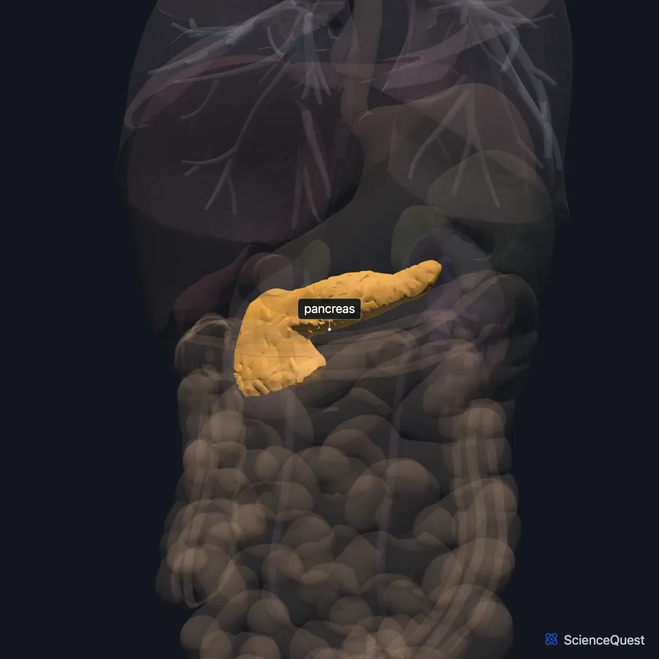
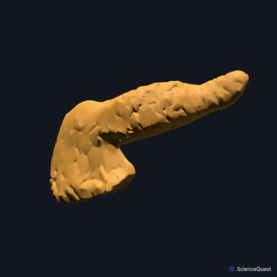
Where is the pancreas?
It lies across the upper abdomen behind the peritoneum, over the first and second lumbar vertebrae, between the duodenum on the right and the spleen on the left. (StatPearls, Pancreas)
Its head sits in the C-shaped curve of the duodenum, on the inferior vena cava and renal vein. Its body crosses the aorta and the second lumbar vertebra, and its tail tapers into the hilum of the spleen. (StatPearls, Pancreas; OpenStax A&P 2e, 23.6)
What are the parts of the pancreas?
It has a head, neck, body and tail. On its way to the duodenum the bile duct runs in a groove on the back of the head, or through the gland itself. (StatPearls, Pancreas)
About 80 percent of it is exocrine tissue, clusters of acinar cells called acini that release enzymes into small ducts. These feed the main pancreatic duct, which joins the bile duct at the hepatopancreatic ampulla and opens into the descending duodenum. (StatPearls, Pancreas; OpenStax A&P 2e, 23.6)
Islets of Langerhans make up as much as 2 percent of the pancreas. Beta cells make insulin, alpha cells glucagon, delta cells somatostatin and PP cells pancreatic polypeptide. (OpenStax A&P 2e, 23.6 and 17.9; StatPearls, Physiology, Islets of Langerhans and Pancreas)
It grows from a dorsal and a ventral bud of the foregut. The ventral bud swings round the duodenum, fuses with the dorsal bud and forms the lower head and uncinate process; the dorsal bud forms the upper head, body and tail. (StatPearls, Pancreas)
What does the pancreas do?
It makes enzymes that digest starch, proteins and fats, among them amylase, lipase and trypsinogen. It also secretes a bicarbonate-rich fluid that neutralizes acid chyme arriving from the stomach. (OpenStax A&P 2e, 23.6; StatPearls, Physiology, Digestion and Physiology, Secretin)
Its protein-digesting enzymes leave in inactive forms. In the duodenum, enterokinase (enteropeptidase) turns trypsinogen into trypsin, which activates the rest. Secreted in active form, they would digest the gland itself, as happens in pancreatitis. (OpenStax A&P 2e, 23.6; StatPearls, Physiology, Digestion)
Acid chyme in the duodenum releases secretin, which makes the pancreas pour out bicarbonate and water. CCK, also from the duodenum, acts on the acinar cells and triggers the release of digestive enzymes. (OpenStax A&P 2e, 23.6; StatPearls, Physiology, Secretin and Pancreas)
Insulin, glucagon and the other islet hormones work together to control blood glucose. (OpenStax A&P 2e, 17.9; StatPearls, Physiology, Pancreas)
What lies around and supplies the pancreas?
Behind its body lie the aorta, the superior mesenteric artery, the left adrenal gland, the left kidney and the renal vessels. Its tail lies in front of the left kidney, close to the splenic hilum and the left colic flexure of the colon. (StatPearls, Pancreas)
Its arteries come from the splenic artery, the superior mesenteric artery and the common hepatic artery. Pancreaticoduodenal arteries feed the head, and the splenic artery feeds the body and tail. (StatPearls, Pancreas)
The head drains to the superior mesenteric vein, the neck and body to the splenic vein, and the two veins join to form the portal vein. (StatPearls, Pancreas)
Secretomotor parasympathetic fibers come from the posterior vagal trunk. Sympathetic fibers come from the splanchnic nerves of T6 to T10 and the celiac plexus. (StatPearls, Pancreas)
How does the pancreas vary?
Pancreas divisum, where the two buds fail to fuse, is its most common developmental anomaly, found in about 10 to 15 percent of people. Annular pancreas comes from incomplete rotation of the ventral bud and can block the duodenum in infancy. (StatPearls, Pancreas)
Where the sources differ
Sources differ on the mix of islet cells: OpenStax puts beta cells at about 75 percent of an islet and alpha cells at about 20, while StatPearls gives up to 60 and up to 30 percent. (OpenStax A&P 2e, 17.9; StatPearls, Physiology, Islets of Langerhans)
The sources describe the accessory duct differently. OpenStax says it runs from the pancreas directly into the duodenum, about an inch above the hepatopancreatic ampulla; StatPearls says it usually empties through the ampulla and opens on its own in about 5 percent of people. Elsewhere the same StatPearls article has pancreatic juice entering the duodenum through both the main and accessory ducts, and the StatPearls duodenum article gives the duodenum both a major and a minor papilla. (OpenStax A&P 2e, 23.6; StatPearls, Pancreas and Duodenum)
| Feature | Detail and source |
|---|---|
| Parts | Head, neck, body, tail (StatPearls, Pancreas) |
| Level | Across L1 and L2, behind the peritoneum (StatPearls, Pancreas) |
| Ducts | Main pancreatic duct, to the hepatopancreatic ampulla; accessory duct (StatPearls, Pancreas) |
| Tissue | About 80% exocrine acini; endocrine islets of Langerhans (StatPearls, Pancreas) |
| Arteries | Splenic, superior mesenteric and common hepatic arteries (StatPearls, Pancreas) |
| Nerves | Posterior vagal trunk; splanchnic nerves T6 to T10 and the celiac plexus (StatPearls, Pancreas) |
Touches in this model
Measured on the BodyParts3D meshes: surfaces within 2 mm of the pancreas, nearest first, 12 of 13.
- Duodenum in contact
- Jejunum in contact
- Pancreatic duct in contact
- Celiac artery in contact
- Common hepatic artery in contact
- Inferior vena cava in contact
- Left gastric artery in contact
- Splenic artery in contact
- Splenic vein in contact
- Superior mesenteric artery in contact
- Superior mesenteric vein in contact
- Stomach 0.6 mm away
Sources
- OpenStax Anatomy and Physiology 2e, section 17.9, The Endocrine Pancreas. CC BY-NC-SA 4.0. Read 2026-10-25.
- StatPearls, Anatomy, Abdomen and Pelvis, Pancreas. Saurabh S. Talathi; Adekunle E. Omole; Michael Young. Last update December 13, 2025. CC BY-NC-ND 4.0. Read 2026-10-25.
- OpenStax Anatomy and Physiology 2e, section 23.6, Accessory Organs in Digestion: The Liver, Pancreas, and Gallbladder. CC BY-NC-SA 4.0. Read 2026-10-25.
- StatPearls, Physiology, Islets of Langerhans. Zainab Shahid; Gurdeep Singh. Last update October 3, 2022. CC BY-NC-ND 4.0. Read 2026-10-25.
- StatPearls, Physiology, Digestion. Justin J. Patricia; Amit S. Dhamoon. Last update September 12, 2022. CC BY-NC-ND 4.0. Read 2026-10-25.
- StatPearls, Physiology, Secretin. Nicholas DiGregorio; Sandeep Sharma. Last update May 1, 2023. CC BY-NC-ND 4.0. Read 2026-10-25.
- StatPearls, Physiology, Pancreas. Suzan A. Kamel-ElSayed; Sandeep Mukherjee. Last update May 1, 2023. CC BY-NC-ND 4.0. Read 2026-10-25.
- StatPearls, Anatomy, Abdomen and Pelvis: Duodenum. Adekunle E. Omole; Shekhar Gogna; Arshia Khorasani-Zadeh. Last update January 31, 2026. CC BY-NC-ND 4.0. Read 2026-10-25.
The values are facts and free to use; this page’s selection and presentation are © 2026 ScienceQuest.