Gallbladder
The gallbladder is a small, pear-shaped sac that stores and concentrates bile. When CCK stimulates it, it contracts and pushes bile through the cystic duct and common bile duct into the duodenum. (OpenStax A&P 2e, 23.6; StatPearls, Physiology, Gallbladder and Physiology, Bile Secretion)
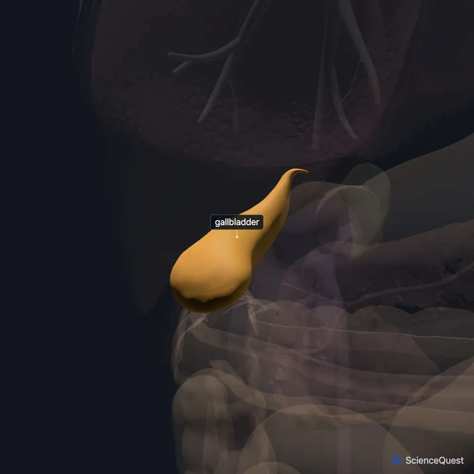
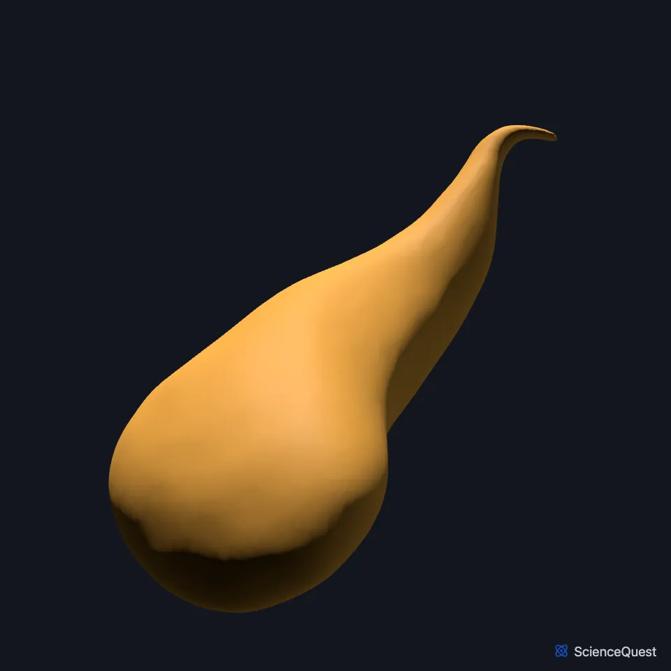
Where is the gallbladder?
It sits in the right upper quadrant of the abdomen. OpenStax places it in a shallow area on the posterior aspect of the right lobe of the liver; StatPearls places it in a fossa between hepatic segments IV and V, fixed to the liver’s visceral surface by connective tissue. (StatPearls, Gallbladder; OpenStax A&P 2e, 23.6)
Visceral peritoneum covers its free surface and forms its outer serosal coat. (OpenStax A&P 2e, 23.6; StatPearls, Gallbladder and Physiology, Gallbladder)
What are the parts of the gallbladder?
It is pear-shaped, with three regions: the wide fundus, the body, and the neck, which angles slightly up toward the hepatic duct. The cystic duct turns down to bridge the neck and the hepatic duct. (StatPearls, Gallbladder; OpenStax A&P 2e, 23.6)
Spiral folds inside the cystic duct, the valves of Heister, adjust the flow of bile. (StatPearls, Gallbladder)
Its lining is simple columnar epithelium folded into rugae, like the stomach’s. Unlike most of the gut, its wall has no submucosa, and a middle coat of smooth muscle contracts to empty it. (OpenStax A&P 2e, 23.6; StatPearls, Gallbladder)
What does the gallbladder do?
It holds 30 to 50 mL of bile. Its lining absorbs water and ions from the bile and concentrates it up to 10-fold. (StatPearls, Gallbladder; OpenStax A&P 2e, 23.6)
Between meals the sphincter of Oddi, the smooth muscle around the hepatopancreatic ampulla, holds the ampulla closed. Bile from the liver then turns back into the gallbladder, which concentrates and stores it until the next meal. (OpenStax A&P 2e, 23.6; StatPearls, Gallbladder)
When the duodenum detects fatty acids, it releases cholecystokinin (CCK). CCK contracts the gallbladder and relaxes the sphincter of Oddi, so bile flows into the duodenum. (StatPearls, Gallbladder)
What connects to the gallbladder?
The cystic duct joins the common hepatic duct to form the common bile duct. The common bile duct and the main pancreatic duct meet in the hepatopancreatic ampulla, where the sphincter of Oddi controls flow into the duodenum. (StatPearls, Gallbladder)
The cystic artery, usually a branch of the right hepatic artery, supplies it. No single cystic vein drains it; short venules run straight into the liver bed. (StatPearls, Gallbladder)
The hepatocystic triangle, or triangle of Calot, lies between the cystic duct, the common hepatic duct and the underside of the liver. It holds the cystic artery and the cystic lymph node. (StatPearls, Gallbladder)
Its nerves arrive through the hepatic plexus: parasympathetic branches of the vagus and sympathetic fibers from the celiac plexus. Irritation of the peritoneum on the diaphragm can refer pain to the right shoulder through the right phrenic nerve. (StatPearls, Gallbladder)
Its neck is sometimes joined to the first part of the duodenum by loose fibrous bands, the cholecystoduodenal fold. (StatPearls, Gallbladder)
How do gallstones form?
High cholesterol, hemolytic disorders and biliary infection change the make-up of bile, which can then crystallize into gallstones. Cholesterol stones are the most common type. (StatPearls, Gallbladder)
Where the sources differ
Its length differs slightly by source: 8 to 10 cm in OpenStax, 7 to 10 cm, with a width of 4 cm, in StatPearls. (OpenStax A&P 2e, 23.6; StatPearls, Gallbladder)
| Feature | Detail and source |
|---|---|
| Parts | Fundus, body, neck (OpenStax A&P 2e, 23.6; StatPearls, Gallbladder) |
| Holds | 30 to 50 mL of bile (StatPearls, Gallbladder) |
| Duct | Cystic duct, which joins the common hepatic duct to form the common bile duct (StatPearls, Gallbladder) |
| Artery | Cystic artery, usually from the right hepatic artery (StatPearls, Gallbladder) |
| Nerves | Hepatic plexus: vagus and celiac plexus fibers (StatPearls, Gallbladder) |
Touches in this model
Measured on the BodyParts3D meshes: surfaces within 2 mm of the gallbladder, nearest first.
Sources
- OpenStax Anatomy and Physiology 2e, section 23.6, Accessory Organs in Digestion: The Liver, Pancreas, and Gallbladder. CC BY-NC-SA 4.0. Read 2026-10-25.
- StatPearls, Physiology, Gallbladder. Mark W. Jones; Sarang Kashyap; Jeffrey G. Deppen. Last update May 1, 2023. CC BY-NC-ND 4.0. Read 2026-10-25.
- StatPearls, Physiology, Bile Secretion. Melanie Hundt; Hajira Basit; Savio John. Last update September 26, 2022. CC BY-NC-ND 4.0. Read 2026-10-25.
- StatPearls, Anatomy, Abdomen and Pelvis: Gallbladder. Mark W. Jones; Bradley Boget; Tafline C. Arbor; Michael Young. Last update July 5, 2026. CC BY-NC-ND 4.0. Read 2026-10-25.
The values are facts and free to use; this page’s selection and presentation are © 2026 ScienceQuest.