Duodenum
The duodenum is the first and shortest part of the small intestine, a C-shaped segment that runs from the pylorus of the stomach to the jejunum. (StatPearls, Duodenum; OpenStax A&P 2e, 23.5)
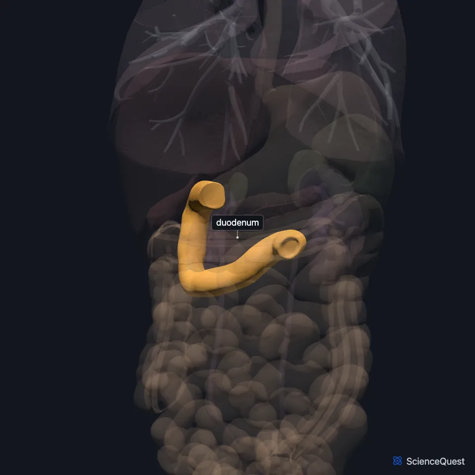
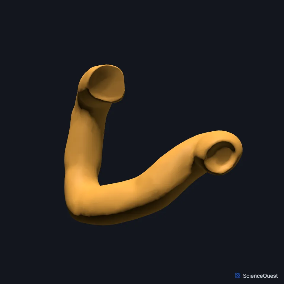
Where is the duodenum?
After its first part it lies behind the peritoneum and curves in a C around the head of the pancreas. At the duodenojejunal junction the gut comes back inside the peritoneum as the jejunum. (OpenStax A&P 2e, 23.5; StatPearls, Duodenum and Small Intestine)
Only the first 2 cm of its superior part is mobile, with a mesentery, and lies inside the peritoneum. The rest is retroperitoneal. (StatPearls, Small Intestine)
What are the four parts of the duodenum?
The first part, the duodenal bulb, is tied to the underside of the liver by the hepatoduodenal ligament, which encloses the portal vein, the hepatic artery and the common bile duct. (StatPearls, Duodenum)
The second, descending part lies in front of the inferior vena cava and the right kidney, with the head of the pancreas in its curve. The third crosses from right to left in front of the aorta and inferior vena cava, with the superior mesenteric vessels in front of it, and the fourth rises to become the jejunum. (StatPearls, Duodenum)
Duodenal (Brunner’s) glands in its submucosa set it apart from the rest of the small intestine. They secrete mucus and bicarbonate that buffer acid chyme from the stomach. (OpenStax A&P 2e, 23.5; StatPearls, Physiology, Small Bowel and Physiology, Secretin)
What does the duodenum do?
Bile and pancreatic juice enter it at the major duodenal papilla in its second part. The bile duct and the main pancreatic duct join in the hepatopancreatic ampulla, and the sphincter of Oddi controls flow from the ampulla into the duodenum. (OpenStax A&P 2e, 23.5; StatPearls, Duodenum)
Acid chyme makes its lining release secretin, which brings in water and bicarbonate to neutralize the acid. Fatty acids and amino acids release cholecystokinin, which slows stomach emptying, contracts the gallbladder and relaxes the sphincter of Oddi. (StatPearls, Duodenum)
It also sets the pace of stomach emptying. Fat arriving in the duodenum releases CCK, which slows emptying and gives fats more time to be digested. (OpenStax A&P 2e, 23.4; StatPearls, Physiology, Small Bowel and Physiology, Bile)
Segmentation, the back-and-forth mixing of chyme, is fastest here, at about 12 contractions a minute. With the upper jejunum, the duodenum is the main site where dietary iron is absorbed. (OpenStax A&P 2e, 23.5; StatPearls, Small Intestine)
What supplies and holds the duodenum?
It shares its blood supply with the head of the pancreas. The superior pancreaticoduodenal artery, from the gastroduodenal artery, feeds the upper part, and the inferior pancreaticoduodenal artery, from the superior mesenteric artery, feeds the lower part. (StatPearls, Duodenum)
It develops from the end of the foregut and the start of the midgut. The celiac artery supplies the foregut and the superior mesenteric artery the midgut. (StatPearls, Small Intestine)
At the duodenojejunal flexure, the ligament of Treitz tethers the gut to the posterior wall. It marks the boundary between the upper and lower gastrointestinal tract. (StatPearls, Small Intestine)
Branches of the vagal trunks give it parasympathetic fibers. Sympathetic fibers from spinal segments T5 to T9 travel in the greater splanchnic nerve to the celiac ganglia. (StatPearls, Duodenum)
Where can the duodenum be compressed or ulcerated?
In superior mesenteric artery syndrome, the third or fourth part of the duodenum is compressed between the superior mesenteric artery and the aorta. (StatPearls, Duodenum)
Duodenal ulcers form most often on the back wall of the first part. An ulcer there can injure the gastroduodenal artery. (StatPearls, Small Intestine and Duodenum)
Where the sources differ
Its length differs by source: 25.4 cm (10 in) in OpenStax, 25 to 30 cm in the StatPearls duodenum article, and 20 to 25 cm in the StatPearls small intestine article. (OpenStax A&P 2e, 23.5; StatPearls, Duodenum and Small Intestine)
They also disagree on how much of the duodenum lies inside the peritoneum. The StatPearls small intestine article says only the first 2 cm of the superior part, and the StatPearls duodenum article the whole first segment; OpenStax has the duodenum turning retroperitoneal just past the pyloric sphincter. (StatPearls, Small Intestine and Duodenum; OpenStax A&P 2e, 23.5)
| Feature | Detail and source |
|---|---|
| Parts | Superior (duodenal bulb), descending, horizontal, ascending (OpenStax A&P 2e, 23.5; StatPearls, Duodenum) |
| Openings | Major and minor duodenal papillae, in the descending part (StatPearls, Duodenum) |
| Peritoneum | Retroperitoneal beyond the first 2 cm of the superior part (StatPearls, Small Intestine) |
| Arteries | Superior and inferior pancreaticoduodenal arteries (StatPearls, Duodenum) |
| Ends | Duodenojejunal flexure, held by the ligament of Treitz (StatPearls, Small Intestine) |
Touches in this model
Measured on the BodyParts3D meshes: surfaces within 2 mm of the duodenum, nearest first.
- Colon in contact
- Gallbladder in contact
- Jejunum in contact
- Liver in contact
- Mesocolic taenia in contact
- Pancreas in contact
- Pancreatic duct in contact
- Stomach in contact
- Inferior vena cava 1.3 mm away
- Descending aorta 1.9 mm away
Sources
- StatPearls, Anatomy, Abdomen and Pelvis: Duodenum. Adekunle E. Omole; Shekhar Gogna; Arshia Khorasani-Zadeh. Last update January 31, 2026. CC BY-NC-ND 4.0. Read 2026-10-25.
- OpenStax Anatomy and Physiology 2e, section 23.5, The Small and Large Intestines. CC BY-NC-SA 4.0. Read 2026-10-25.
- StatPearls, Anatomy, Abdomen and Pelvis, Small Intestine. Jason T. Collins; Amanda Nguyen; Adekunle E. Omole; Madhu Badireddy. Last update February 18, 2025. CC BY-NC-ND 4.0. Read 2026-10-25.
- StatPearls, Physiology, Small Bowel. Elizabeth M. Fish; Karlie R. Shumway; Bracken Burns. Last update January 31, 2024. CC BY-NC-ND 4.0. Read 2026-10-25.
- StatPearls, Physiology, Secretin. Nicholas DiGregorio; Sandeep Sharma. Last update May 1, 2023. CC BY-NC-ND 4.0. Read 2026-10-25.
- OpenStax Anatomy and Physiology 2e, section 23.4, The Stomach. CC BY-NC-SA 4.0. Read 2026-10-25.
- StatPearls, Physiology, Bile. Ali N. Almajid; Kavin Sugumar. Last update September 12, 2022. CC BY-NC-ND 4.0. Read 2026-10-25.
The values are facts and free to use; this page’s selection and presentation are © 2026 ScienceQuest.