Liver
The liver is the largest gland in the body, about 2 percent of adult body weight. Besides making bile, it takes up absorbed nutrients and clears drugs and other harmful substances. (OpenStax A&P 2e, 23.6; StatPearls, Liver and Physiology, Liver)
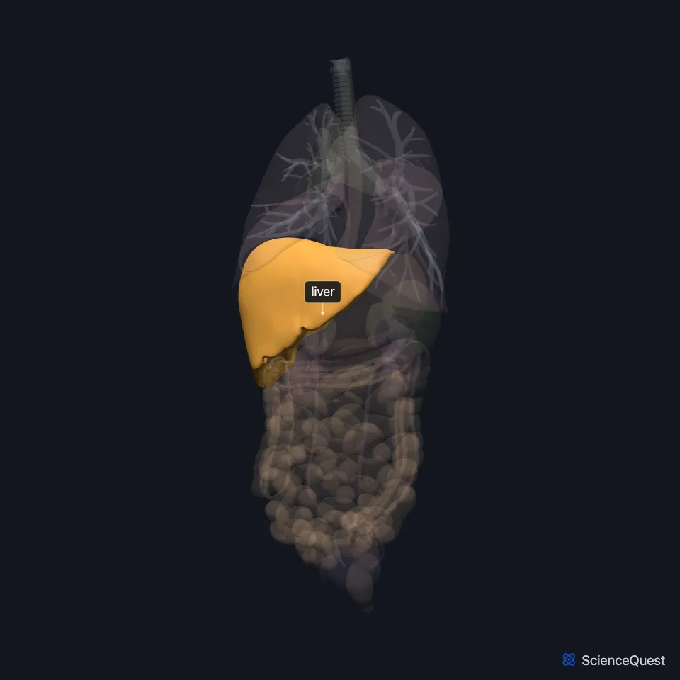
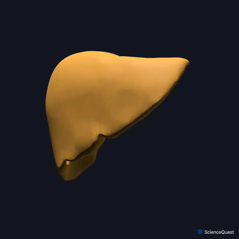
Where is the liver?
It lies below the diaphragm in the right upper quadrant of the abdomen, where the ribs protect it. It reaches from the fifth intercostal space in the midclavicular line to the right costal margin. (OpenStax A&P 2e, 23.6; StatPearls, Liver)
Peritoneum covers most of it. A bare area on its upper back surface, where the diaphragm and inferior vena cava lie, has none, and the coronary ligament forms at its border. (StatPearls, Liver)
Its underside lies close to the gallbladder and the right kidney. (StatPearls, Liver)
What are the lobes of the liver?
On its front surface the falciform ligament divides it into right and left. The free edge of the ligament carries the round ligament, the remnant of the umbilical vein. (StatPearls, Liver)
The caudate lobe lies behind, bounded by the ligamentum venosum, the inferior vena cava and the porta hepatis. The quadrate lobe lies below, in front of the porta hepatis and next to the gallbladder. (StatPearls, Liver; OpenStax A&P 2e, 23.6)
Its 8 functional subdivisions follow the blood supply and do not match the anatomical lobes. (StatPearls, Liver)
Its working unit is the lobule, a hexagon of hepatocytes around a central vein. At its corners sit portal triads, each a branch of the bile duct, the portal vein and the hepatic artery. (StatPearls, Liver)
Kupffer cells, the liver’s resident macrophages, sit in its sinusoids and filter unwanted or harmful material out of the blood. (OpenStax A&P 2e, 23.6; StatPearls, Liver and Physiology, Liver)
What does the liver do?
Hepatocytes make bile, which flows to the duodenum. There, bile salts emulsify fat, breaking large droplets into small ones that digestive enzymes can reach. (OpenStax A&P 2e, 23.6; StatPearls, Physiology, Bile Secretion and Physiology, Bile)
It makes proteins such as albumin, fibrinogen and prothrombin, stores glycogen, makes new glucose (gluconeogenesis) and converts ammonia to urea. (StatPearls, Liver)
It processes toxic substances absorbed from the gut, and it stores vitamins and minerals such as iron. (StatPearls, Liver)
Old red cells broken down in the spleen yield bilirubin, which travels to the liver bound to albumin and leaves in the bile. (OpenStax A&P 2e, 23.6; StatPearls, Physiology, Bile Secretion and Physiology, Spleen)
What supplies the liver?
Blood reaches it from two sources: 75 to 80 percent of the volume through the portal vein and 20 to 25 percent through the hepatic artery. Both enter at the porta hepatis. (StatPearls, Liver; OpenStax A&P 2e, 23.6)
About 75 percent of the liver’s blood arrives in the portal vein, which carries absorbed nutrients from the gut, and about 25 percent in the hepatic artery. Because it receives blood from so much of the body, most cancers in the liver have spread there from elsewhere. (OpenStax A&P 2e, 23.6; StatPearls, Physiology, Liver and Liver)
Blood leaves through the left, middle and right hepatic veins into the inferior vena cava. (StatPearls, Liver; OpenStax A&P 2e, 23.6)
Bile leaves through the right and left hepatic ducts, which join outside the liver as the common hepatic duct. That joins the cystic duct from the gallbladder to form the common bile duct, which empties into the duodenum. (OpenStax A&P 2e, 23.6; StatPearls, Gallbladder, Physiology, Bile Secretion and Duodenum)
The lesser omentum ties it to the lesser curvature of the stomach. Its nerves come through the hepatic plexus, with sympathetic fibers from the celiac plexus and parasympathetic fibers from the vagal trunks. (OpenStax A&P 2e, 23.6; StatPearls, Liver)
Where the sources differ
The sources place the Kupffer cells differently. OpenStax and the StatPearls liver anatomy chapter put them in the sinusoids, while the StatPearls liver physiology chapter places them in the space of Disse, between the sinusoid lining and the hepatocytes. (OpenStax A&P 2e, 23.6; StatPearls, Liver and Physiology, Liver)
Estimates of daily bile output differ: OpenStax gives about a liter, and StatPearls about 600 mL. (OpenStax A&P 2e, 23.6; StatPearls, Physiology, Bile Secretion)
The sources give bile different colors: StatPearls says bilirubin makes it yellow, while OpenStax says bilirubin colors it green. (StatPearls, Physiology, Bile Secretion; OpenStax A&P 2e, 23.6)
The sources count the lobes differently. OpenStax describes two primary lobes, a large right and a much smaller left, and says some anatomists also identify quadrate and caudate lobes; StatPearls lists four: right, left, caudate and quadrate. (OpenStax A&P 2e, 23.6; StatPearls, Liver)
| Feature | Detail and source |
|---|---|
| Lobes | Right and left; caudate and quadrate counted as lobes by some sources (OpenStax A&P 2e, 23.6; StatPearls, Liver) |
| Blood in | Portal vein (75 to 80%) and hepatic artery (20 to 25%), at the porta hepatis (StatPearls, Liver; OpenStax A&P 2e, 23.6) |
| Blood out | Hepatic veins to the inferior vena cava (OpenStax A&P 2e, 23.6; StatPearls, Liver; Gray’s Anatomy (1918), The Veins) |
| Bile out | Right and left hepatic ducts, then the common hepatic duct (OpenStax A&P 2e, 23.6; StatPearls, Gallbladder) |
| Nerves | Hepatic plexus: celiac plexus and vagal trunks (StatPearls, Liver) |
Touches in this model
Measured on the BodyParts3D meshes: surfaces within 2 mm of the liver, nearest first, 12 of 20.
- Colon in contact
- Duodenum in contact
- Gallbladder in contact
- Lower lobe of right lung in contact
- Right adrenal gland in contact
- Right kidney in contact
- Stomach in contact
- Diaphragm in contact
- Inferior vena cava in contact
- Wall of heart in contact
- Right transversus abdominis 0.2 mm away
- Skin 0.4 mm away
Sources
- OpenStax Anatomy and Physiology 2e, section 23.6, Accessory Organs in Digestion: The Liver, Pancreas, and Gallbladder. CC BY-NC-SA 4.0. Read 2026-10-25.
- StatPearls, Anatomy, Abdomen and Pelvis: Liver. Hamilton Vernon; Chase J. Wehrle; Valentine Sampson K. Alia; Anup Kasi. Last update November 26, 2022. CC BY-NC-ND 4.0. Read 2026-10-25.
- StatPearls, Physiology, Liver. Arjun Kalra; Ekrem Yetiskul; Chase J. Wehrle; Faiz Tuma. Last update May 1, 2023. CC BY-NC-ND 4.0. Read 2026-10-25.
- StatPearls, Physiology, Bile Secretion. Melanie Hundt; Hajira Basit; Savio John. Last update September 26, 2022. CC BY-NC-ND 4.0. Read 2026-10-25.
- StatPearls, Physiology, Bile. Ali N. Almajid; Kavin Sugumar. Last update September 12, 2022. CC BY-NC-ND 4.0. Read 2026-10-25.
- StatPearls, Physiology, Spleen. Vaishali Kapila; Chase J. Wehrle; Faiz Tuma. Last update May 1, 2023. CC BY-NC-ND 4.0. Read 2026-10-25.
- StatPearls, Anatomy, Abdomen and Pelvis: Gallbladder. Mark W. Jones; Bradley Boget; Tafline C. Arbor; Michael Young. Last update July 5, 2026. CC BY-NC-ND 4.0. Read 2026-10-25.
- StatPearls, Anatomy, Abdomen and Pelvis: Duodenum. Adekunle E. Omole; Shekhar Gogna; Arshia Khorasani-Zadeh. Last update January 31, 2026. CC BY-NC-ND 4.0. Read 2026-10-25.
- Gray’s Anatomy of the Human Body, 20th US edition (1918), Angiology, The Veins, page 640. Henry Gray; edited by Warren H. Lewis. Public domain. Read 2026-10-25.
The values are facts and free to use; this page’s selection and presentation are © 2026 ScienceQuest.