Wall of heart
The wall of the heart has three layers of unequal thickness. From the outside in, they are the epicardium, the myocardium and the endocardium. (OpenStax A&P 2e, 19.1; StatPearls, Histology, Heart and Heart and Pericardial Cavity)
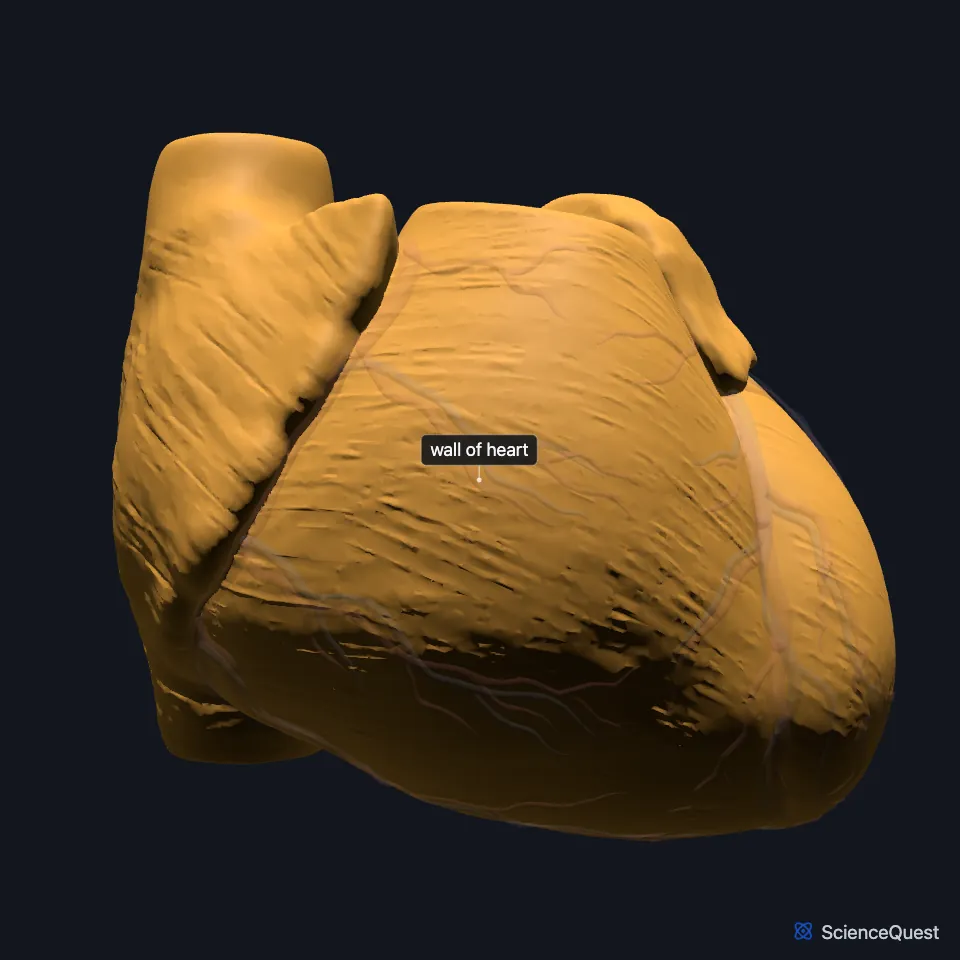
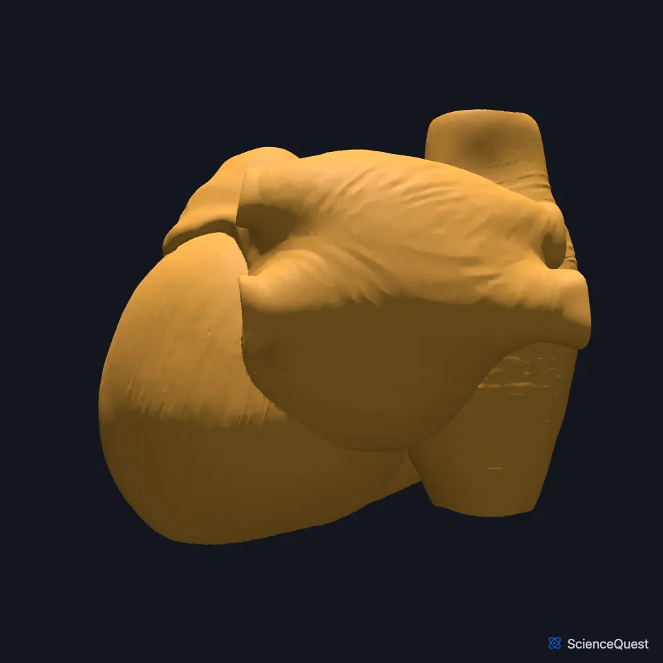
Where is the heart wall?
The heart lies in the mediastinum, between the lungs, inside its own sac, the pericardium. It sits obliquely behind the sternum and the rib cartilages, and the esophagus, aorta and thoracic duct separate its back from the fifth to eighth thoracic vertebrae. (OpenStax A&P 2e, 19.1; Gray 1918, 4b. The Heart)
The heart spans the third to sixth costal cartilages. OpenStax calls its top surface, where the great vessels attach, the base and places it at the third costal cartilage, while Gray’s Anatomy (1918) describes the base as facing upward, backward and to the right and formed mainly by the left atrium. OpenStax places the apex, the lower tip, just left of the sternum between the fourth and fifth ribs; Gray’s puts it behind the fifth left intercostal space, 8 to 9 cm from the midline of the sternum. (OpenStax A&P 2e, 19.1; StatPearls, Heart and Pericardial Cavity; Gray 1918, 4b. The Heart)
A typical adult heart is about 12 cm long, 8 to 9 cm wide at its broadest and 6 cm thick. (OpenStax A&P 2e, 19.1; Gray 1918, 4b. The Heart)
What are the layers of the heart wall?
The epicardium, the outer layer, is the visceral layer of the serous pericardium. Its mesothelial cells secrete pericardial fluid, which lubricates the layers and reduces friction with each heartbeat. (OpenStax A&P 2e, 19.1; StatPearls, Pericardium and Heart and Pericardial Cavity)
The myocardium, the middle layer, is the thickest, especially in the left ventricle. It is made mainly of cardiac muscle cells and connective tissue, and its contractions eject blood from the heart. (OpenStax A&P 2e, 19.1; StatPearls, Heart and Pericardial Cavity and Histology, Heart)
Its muscle fibers wind around the chambers in layers. In the atria, deep looped fibers arch over each atrium from its atrioventricular ring, and deep ring-shaped fibers circle the auricles, the openings of the veins and the fossa ovalis; in the ventricles superficial fibers sweep around the apex. OpenStax describes the overall pattern as two figure 8s, one around the atria and great vessels and a deeper one around the ventricles. (OpenStax A&P 2e, 19.1; Gray 1918, 4b. The Heart)
The endocardium lines the chambers and covers the valves. It is a simple squamous epithelium, the endothelium, continuous with the lining of the blood vessels. (OpenStax A&P 2e, 19.1; StatPearls, Physiology, Cardiac Muscle; Gray 1918, 4b. The Heart)
Cardiac muscle cells branch, and intercalated discs join adjoining cells end to end. Gap junctions in the discs let the electrical signal spread from cell to cell, so the cells contract together. (OpenStax A&P 2e, 19.2; StatPearls, Cardiac Muscle)
What does the heart wall do?
The left ventricle’s wall is about three times as thick as the right’s. It drives blood around the long systemic circuit at far higher pressure, peaking near 120 mmHg against about 25 mmHg in the right ventricle. (OpenStax A&P 2e, 19.1; Gray 1918, 4b. The Heart; StatPearls, Cardiac Cycle)
Septa divide the chambers. Most of the interventricular septum is thick muscle, apart from a thin fibrous upper part, the membranous septum. (OpenStax A&P 2e, 19.1; Gray 1918, 4b. The Heart)
A cardiac muscle action potential lasts about 200 ms and has a plateau phase, held up by calcium flowing into the cell, that skeletal muscle lacks. Relaxation is an active step that lets the heart refill between beats. (OpenStax A&P 2e, 19.2; StatPearls, Physiology, Cardiac Muscle and Cardiac Muscle)
What lies on and around the heart wall?
Fat-filled grooves on its surface, the sulci, carry the major coronary vessels. The coronary sulcus runs between the atria and the ventricles, and two interventricular sulci run between the ventricles. (OpenStax A&P 2e, 19.1; Gray 1918, 4b. The Heart; StatPearls, Heart and Pericardial Cavity)
The coronary arteries supply the myocardium. The left and right coronary arteries both arise from the aorta, just above the aortic valve. (OpenStax A&P 2e, 19.1; StatPearls, Heart and Pericardial Cavity)
The pericardial cavity, filled with serous fluid, lies between the epicardium (the visceral pericardium) and the parietal pericardium. The fibrous pericardium outside it attaches to the sternum and the diaphragm and limits how far the heart moves. (OpenStax A&P 2e, 19.1; StatPearls, Heart and Pericardial Cavity)
How does the heart wall respond to exercise and injury?
Hearts of athletes can be larger than usual. (OpenStax A&P 2e, 19.1; StatPearls, Heart)
Damaged cardiac muscle cells can do little to repair themselves or replace dead cells. After a heart attack, patches of scar tissue often take the place of the dead cells. (OpenStax A&P 2e, 19.2; StatPearls, Histology, Heart and Cardiac Muscle)
| Feature | Detail and source |
|---|---|
| Outer layer | Epicardium, the visceral pericardium (OpenStax A&P 2e, 19.1; StatPearls, Heart and Pericardial Cavity) |
| Middle layer | Myocardium, the thickest, mainly cardiac muscle cells and connective tissue (OpenStax A&P 2e, 19.1; StatPearls, Heart and Pericardial Cavity and Cardiac Muscle) |
| Inner layer | Endocardium, an endothelium that also covers the valves (OpenStax A&P 2e, 19.1; StatPearls, Physiology, Cardiac Muscle) |
| Cell junctions | Intercalated discs, with desmosomes and gap junctions (OpenStax A&P 2e, 19.2; StatPearls, Cardiac Muscle) |
Touches in this model
Measured on the BodyParts3D meshes: surfaces within 2 mm of the wall of heart, nearest first, 12 of 42.
- Anterior interventricular branch of left coronary artery in contact
- Anterior papillary muscle of right ventricle in contact
- Circumflex branch of left coronary artery in contact
- Coronary sinus in contact
- Great cardiac vein in contact
- Marginal branch of right coronary artery in contact
- Middle cardiac vein in contact
- Mitral valve in contact
- Papillary muscle of left ventricle in contact
- Posterior interventricular branch of right coronary artery in contact
- Posterior papillary muscle of left ventricle in contact
- Posterior papillary muscle of right ventricle in contact
Sources
- OpenStax Anatomy and Physiology 2e, section 19.1, Heart Anatomy. CC BY-NC-SA 4.0. Read 2026-10-25.
- StatPearls, Histology, Heart. Anita Arackal; Khalid Alsayouri. Last update January 2, 2023. CC BY-NC-ND 4.0. Read 2026-10-25.
- StatPearls, Anatomy, Thorax, Heart and Pericardial Cavity. John K. Volpe; Amgad N. Makaryus. Last update July 25, 2023. CC BY-NC-ND 4.0. Read 2026-10-25.
- Gray’s Anatomy of the Human Body, 20th US edition (1918), V. Angiology, 4b. The Heart (1918 terms: longitudinal sulci for interventricular sulci, musculi papillares for papillary muscles). Henry Gray. Public domain. Read 2026-10-25.
- StatPearls, Anatomy, Thorax, Pericardium. Adekunle E. Omole; Ali Nassereddin; Marjorie V. Launico. Last update February 14, 2026. CC BY-NC-ND 4.0. Read 2026-10-25.
- StatPearls, Physiology, Cardiac Muscle. Rashelle Ripa; Tom George; Karlie R. Shumway; Yasar Sattar. Last update July 30, 2023. CC BY-NC-ND 4.0. Read 2026-10-25.
- OpenStax Anatomy and Physiology 2e, section 19.2, Cardiac Muscle and Electrical Activity. CC BY-NC-SA 4.0. Read 2026-10-25.
- StatPearls, Anatomy, Thorax, Cardiac Muscle. Anthony Saxton; Muhammad Ali Tariq; Bruno Bordoni. Last update August 8, 2023. CC BY-NC-ND 4.0. Read 2026-10-25.
- StatPearls, Physiology, Cardiac Cycle. Joshua D. Pollock; Amgad N. Makaryus. Last update October 3, 2022. CC BY-NC-ND 4.0. Read 2026-10-25.
- StatPearls, Anatomy, Thorax, Heart. Ibraheem Rehman; Afzal Rehman. Last update August 28, 2023. CC BY-NC-ND 4.0. Read 2026-10-25.
The values are facts and free to use; this page’s selection and presentation are © 2026 ScienceQuest.