Circumflex branch of left coronary artery
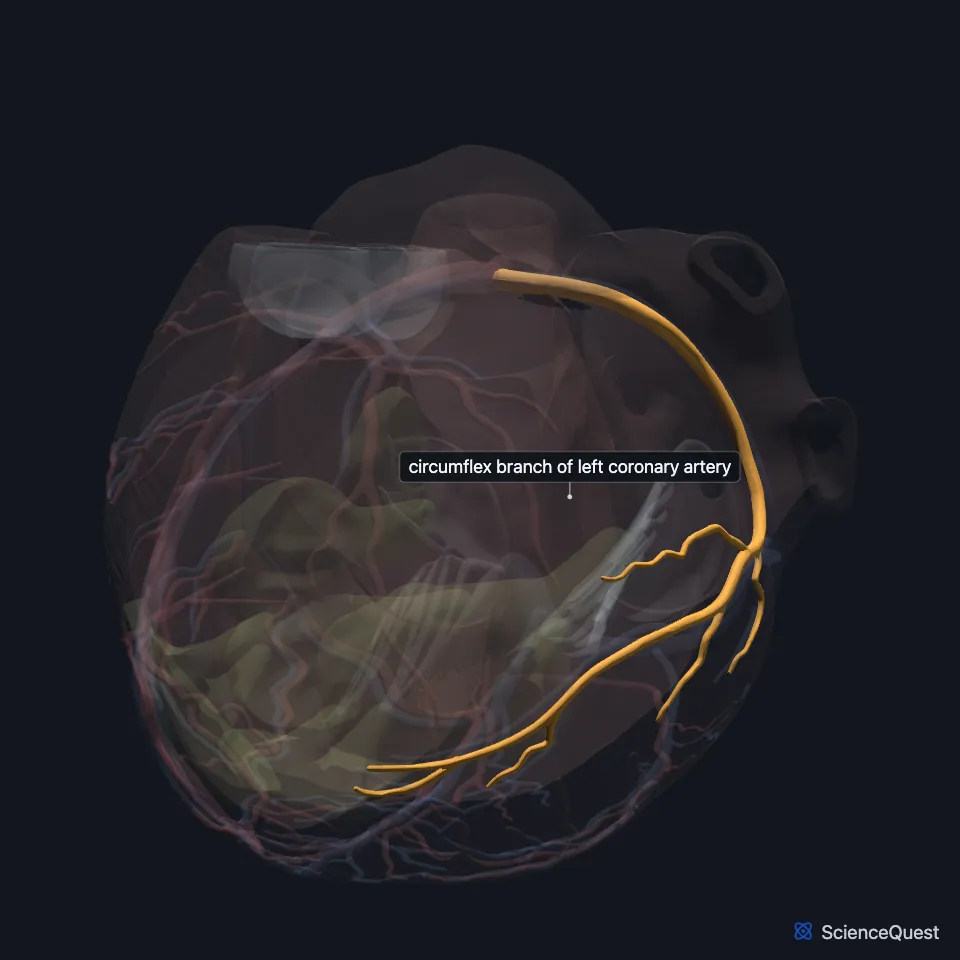
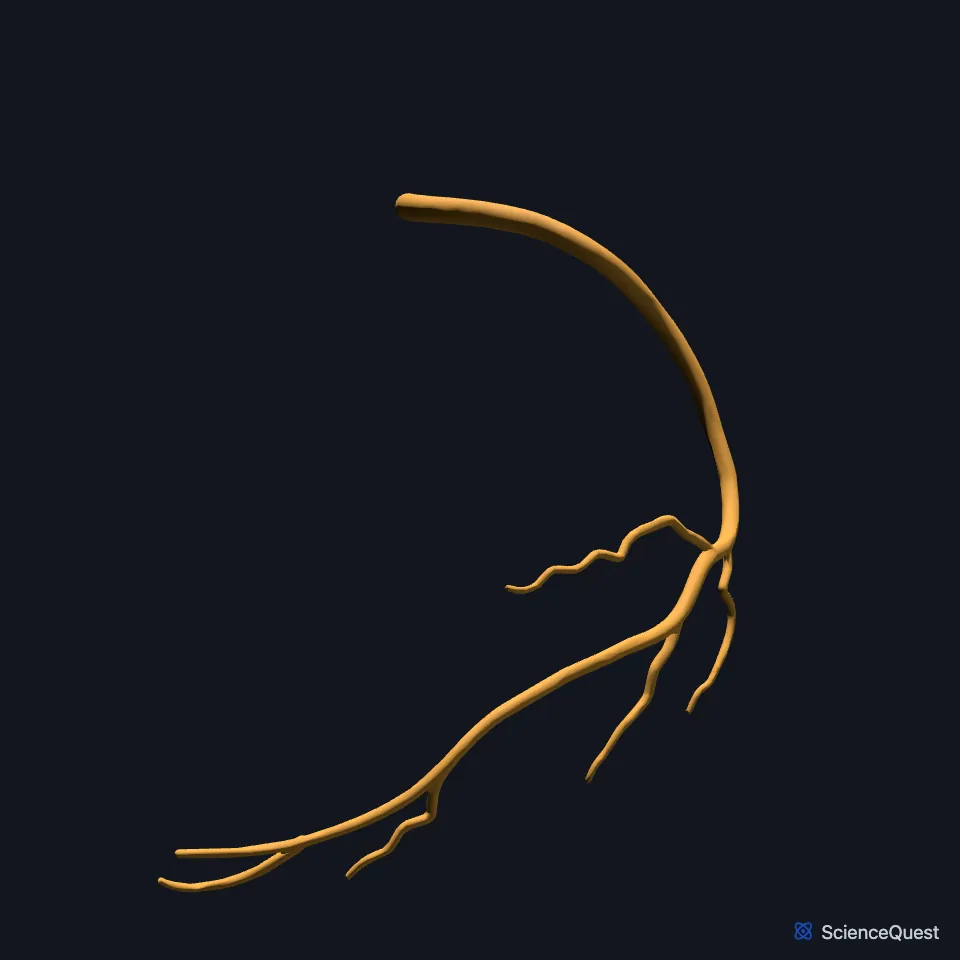
The circumflex artery is a branch of the left coronary artery that follows the coronary sulcus to the left. (OpenStax A&P 2e, 19.1; StatPearls, Heart Arteries; Gray 1918, 2. The Aorta)
Where does the circumflex artery run?
It runs in the left atrioventricular groove, the part of the coronary sulcus between the left atrium and the left ventricle, toward the left side of the heart. (StatPearls, Heart Left Anterior Descending Artery and Heart Arteries)
It and the anterior interventricular artery are the two branches into which the left main coronary artery divides. (StatPearls, Heart Left Anterior Descending Artery)
What are the branches of the circumflex artery?
It divides into the left marginal artery and the posterior left ventricular artery. (StatPearls, Heart Arteries)
What does the circumflex artery supply?
With its branches it supplies the back and side walls of the left ventricle. (StatPearls, Heart Arteries and Heart Left Anterior Descending Artery)
It shares the supply of the anterior papillary muscle of the left ventricle with the anterior interventricular artery. In some hearts its branches also supply the posterior papillary muscle, which the StatPearls article on the papillary muscles calls the posteromedial. (StatPearls, Mitral Valve and Heart Papillary Muscles)
Where does the circumflex artery meet other vessels?
It ends by joining small branches of the right coronary artery in the coronary sulcus. (OpenStax A&P 2e, 19.1; StatPearls, Heart Arteries)
Does the circumflex artery vary?
In some hearts it leaves the aorta separately from the anterior interventricular artery, with no left main stem. That separate origin is the most common congenital anomaly of the coronary arteries. (StatPearls, Heart Left Anterior Descending Artery)
Where the sources differ
The sources pair different veins with this artery. OpenStax says the posterior cardiac vein runs beside its marginal branch and drains the area that branch supplies. StatPearls and Gray’s Anatomy (1918) instead describe two veins here, the left marginal vein along the left margin of the heart and the posterior vein of the left ventricle on its inferior surface, and neither pairs a vein with the marginal branch. (OpenStax A&P 2e, 19.1; StatPearls, Heart Veins; Gray 1918, 3. The Systemic Veins)
| Feature | Detail and source |
|---|---|
| Branch of | Left coronary artery (OpenStax A&P 2e, 19.1; StatPearls, Heart Arteries) |
| Runs in | The left atrioventricular groove of the coronary sulcus (StatPearls, Heart Left Anterior Descending Artery) |
| Branches | Left marginal artery, posterior left ventricular artery (StatPearls, Heart Arteries) |
| Supplies | Back and side walls of the left ventricle (StatPearls, Heart Arteries) |
Touches in this model
Measured on the BodyParts3D meshes: surfaces within 2 mm of the circumflex branch of left coronary artery, nearest first.
- Anterior interventricular branch of left coronary artery in contact
- Great cardiac vein in contact
- Set of posterior veins of left ventricle in contact
- Stem of left coronary artery in contact
- Wall of heart in contact
- Lower lobe of left lung in contact
- Upper lobe of left lung in contact
Sources
- OpenStax Anatomy and Physiology 2e, section 19.1, Heart Anatomy. CC BY-NC-SA 4.0. Read 2026-10-25.
- StatPearls, Anatomy, Thorax, Heart Arteries. Raheel Chaudhry; Sajedur Rahman; Mark A. Law. Last update July 24, 2023. CC BY-NC-ND 4.0. Read 2026-10-25.
- Gray’s Anatomy of the Human Body, 20th US edition (1918), VI. The Arteries, 2. The Aorta (1918 terms: longitudinal sulcus for interventricular sulcus, anterior aortic sinuses for the coronary sinuses of the aorta). Henry Gray. Public domain. Read 2026-10-25.
- StatPearls, Anatomy, Thorax, Heart Left Anterior Descending (LAD) Artery. Himanshu Jindal; Adegbenro O. Fakoya. Last update April 12, 2026. CC BY-NC-ND 4.0. Read 2026-10-25.
- StatPearls, Anatomy, Thorax, Mitral Valve. Felipe Sanchez Vaca; Bruno Bordoni. Last update July 24, 2023. CC BY-NC-ND 4.0. Read 2026-10-25.
- StatPearls, Anatomy, Thorax, Heart Papillary Muscles. Natalie L. Rich; Yusuf S. Khan. Last update July 24, 2023. CC BY-NC-ND 4.0. Read 2026-10-25.
- StatPearls, Anatomy, Thorax, Heart Veins. Julia H. Miao; Amgad N. Makaryus. Last update July 24, 2023. CC BY-NC-ND 4.0. Read 2026-10-25.
- Gray’s Anatomy of the Human Body, 20th US edition (1918), VII. The Veins, 3. The Systemic Veins (1918 terms: longitudinal sulcus for interventricular sulcus). Henry Gray. Public domain. Read 2026-10-25.
The values are facts and free to use; this page’s selection and presentation are © 2026 ScienceQuest.