Coronary sinus
The coronary sinus is the largest vein of the heart and lies on its back. It runs in the atrioventricular sulcus and empties into the right atrium. (OpenStax A&P 2e, 19.1; StatPearls, Coronary Sinus)
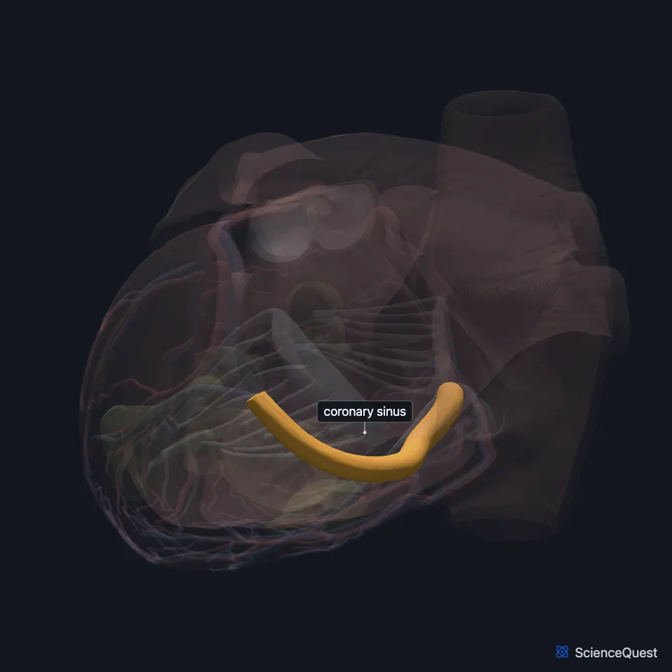
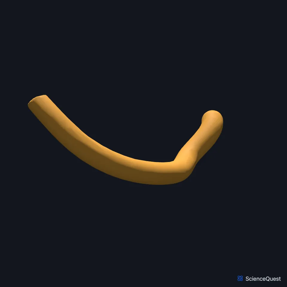
Where is the coronary sinus?
It is the largest coronary vein, 3 to 5 cm long and about 1 cm across. It begins on the back of the heart between the left atrium and the left ventricle and runs along the left posterior atrioventricular groove. (StatPearls, Coronary Sinus)
It opens into the lower back part of the right atrium, just medial to the inferior vena cava and above the septal leaflet of the tricuspid valve. (StatPearls, Coronary Sinus; OpenStax A&P 2e, 19.1)
Which veins drain into the coronary sinus?
It begins where the great cardiac vein meets the oblique vein of the left atrium. Along its course the posterior vein of the left ventricle, the middle cardiac vein and the small cardiac vein drain into it, in that order. (StatPearls, Coronary Sinus)
The tributaries other than the oblique vein of the left atrium have valves where they join, though the form of these valves varies between people. The valve of Vieussens marks where the sinus begins, and the Thebesian valve at its opening stops blood in the right atrium flowing back into it. (StatPearls, Coronary Sinus)
A sleeve of striated cardiac muscle covers it, continuous with the muscle of both atria. (StatPearls, Coronary Sinus)
What does the coronary sinus do?
It drains most of the coronary veins, returning blood from the heart muscle to the right atrium. (OpenStax A&P 2e, 19.1; Gray 1918, 3. The Systemic Veins)
The right atrium receives blood from the two venae cavae and from the coronary sinus. (OpenStax A&P 2e, 19.3; StatPearls, Heart)
It returns about 55% of the heart’s venous blood. The anterior cardiac veins, which bypass it and drain straight into the right atrium, return about 35%. (StatPearls, Coronary Sinus; OpenStax A&P 2e, 19.1)
What lies next to the coronary sinus?
The lip of its opening forms one side of the triangle of Koch, with the tricuspid valve and the tendon of Todaro. The atrioventricular node lies in that triangle. (StatPearls, Heart)
On the back of the heart the posterior vein of the left ventricle runs between the middle and great cardiac veins and drains into the sinus. (StatPearls, Heart Veins)
Does the coronary sinus vary?
In a persistent left superior vena cava, a rare variant, blood from the left arm, head and neck drains into the coronary sinus before it reaches the right atrium. (StatPearls, Coronary Sinus)
Some people have two coronary sinuses. (StatPearls, Heart Veins)
In the embryo, it forms from the left horn of the sinus venosus. (StatPearls, Coronary Sinus)
Where the sources differ
Three StatPearls articles give different splits of the heart’s venous return. The coronary sinus article gives the smallest cardiac veins 10%; the heart veins article gives the smaller venous system a quarter; and the heart arteries article gives the greater cardiac veins 95%. (StatPearls, Coronary Sinus, Heart Veins and Heart Arteries)
| Feature | Detail and source |
|---|---|
| Lies in | The posterior atrioventricular groove (StatPearls, Coronary Sinus) |
| Size | 3 to 5 cm long, about 1 cm across (StatPearls, Coronary Sinus) |
| Tributaries | Great cardiac vein, oblique vein of the left atrium, posterior vein of the left ventricle, middle cardiac vein, small cardiac vein (StatPearls, Coronary Sinus) |
| Empties into | The right atrium, through the Thebesian valve (StatPearls, Coronary Sinus) |
Touches in this model
Measured on the BodyParts3D meshes: surfaces within 2 mm of the coronary sinus, nearest first.
- Great cardiac vein in contact
- Middle cardiac vein in contact
- Set of posterior veins of left ventricle in contact
- Tricuspid valve in contact
- Wall of heart in contact
- Lower lobe of left lung in contact
- Trunk of right coronary artery 1.5 mm away
Sources
- OpenStax Anatomy and Physiology 2e, section 19.1, Heart Anatomy. CC BY-NC-SA 4.0. Read 2026-10-25.
- StatPearls, Anatomy, Thorax, Coronary Sinus. Alexander Wilson; Beenish S. Bhutta. Last update July 24, 2023. CC BY-NC-ND 4.0. Read 2026-10-25.
- Gray’s Anatomy of the Human Body, 20th US edition (1918), VII. The Veins, 3. The Systemic Veins (1918 terms: longitudinal sulcus for interventricular sulcus). Henry Gray. Public domain. Read 2026-10-25.
- OpenStax Anatomy and Physiology 2e, section 19.3, Cardiac Cycle. CC BY-NC-SA 4.0. Read 2026-10-25.
- StatPearls, Anatomy, Thorax, Heart. Ibraheem Rehman; Afzal Rehman. Last update August 28, 2023. CC BY-NC-ND 4.0. Read 2026-10-25.
- StatPearls, Anatomy, Thorax, Heart Veins. Julia H. Miao; Amgad N. Makaryus. Last update July 24, 2023. CC BY-NC-ND 4.0. Read 2026-10-25.
- StatPearls, Anatomy, Thorax, Heart Arteries. Raheel Chaudhry; Sajedur Rahman; Mark A. Law. Last update July 24, 2023. CC BY-NC-ND 4.0. Read 2026-10-25.
The values are facts and free to use; this page’s selection and presentation are © 2026 ScienceQuest.