Tricuspid valve
The tricuspid valve, or right atrioventricular valve, lies between the right atrium and the right ventricle. It usually has three cusps, folds of the heart’s lining strengthened by fibrous tissue. (OpenStax A&P 2e, 19.1; StatPearls, Histology, Heart; Gray 1918, 4b. The Heart; FMA, FMA7234)
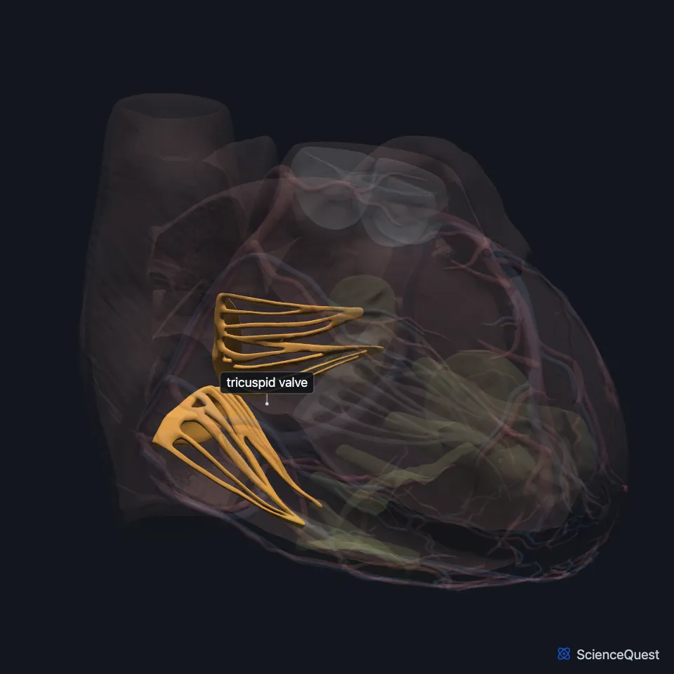
Where is the tricuspid valve?
It is the largest of the heart valves and the one set closest to the apex. Its normal area is 7 to 9 cm². (StatPearls, Tricuspid Valve Repair)
The right atrium above it collects blood from the superior and inferior venae cavae and from the coronary sinus, which drains the heart muscle. (OpenStax A&P 2e, 19.1; StatPearls, Heart)
Its closing is heard loudest in the fourth left intercostal space. (StatPearls, Heart Sounds)
What are the parts of the tricuspid valve?
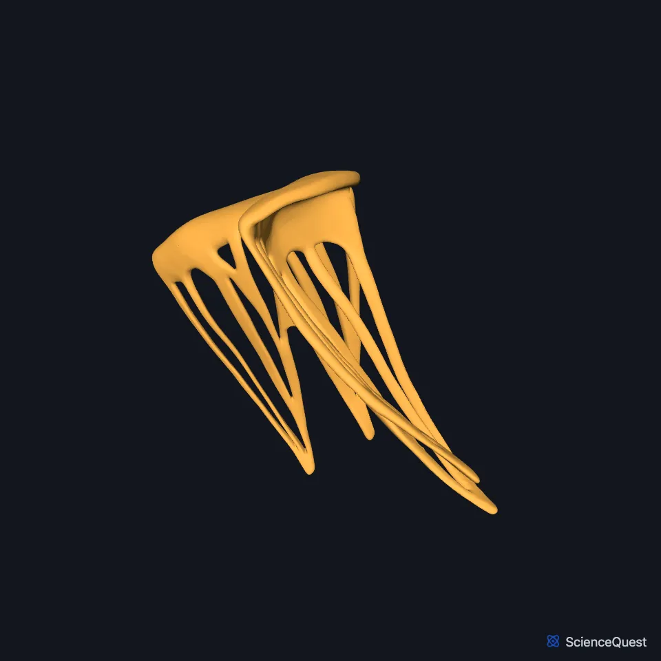
Its three leaflets are the anterior, the posterior and the septal. The anterior is the largest and moves the most, the septal moves the least, and the posterior is the shortest around the ring. (StatPearls, Tricuspid Valve Repair)
Chordae tendineae tie the flaps to papillary muscles in the right ventricle. The cords are made mainly of dense regular connective tissue with collagen and elastic fibers. (OpenStax A&P 2e, 19.1; StatPearls, Histology, Heart)
Its annulus is a D-shaped ring that moves with the heart. The longer, C-shaped part follows the free wall of the right ventricle, and the shorter, straighter part lies along the septum. (StatPearls, Tricuspid Valve Repair)
What does the tricuspid valve do?
While the heart relaxes, blood from the right atrium flows through the open valve into the right ventricle. (OpenStax A&P 2e, 19.3 and 19.1; StatPearls, Heart and Pericardial Cavity)
When the right ventricle contracts, its papillary muscles contract too and pull on the chordae. That stops the flaps from being forced into the atrium and blood from flowing back. (OpenStax A&P 2e, 19.1; Gray 1918, 4b. The Heart)
Its closure, with the mitral valve’s, makes the first heart sound, S1. At rest the two closures come too close together to tell apart. (StatPearls, Heart Sounds)
The mean pressure difference across the valve is less than 2 mm Hg, because the valve is large and pressures on the right side of the heart are low. (StatPearls, Tricuspid Valve Repair)
What lies around the tricuspid valve?
The right ventricle has three papillary muscles. The anterior, the largest, rises from the anterior wall, the posterior from the inferior wall, and the septal, the smallest, from the interventricular septum. (StatPearls, Heart Papillary Muscles)
Its septal leaflet attaches to the septum less than 10 mm nearer the apex than the anterior leaflet of the mitral valve. (StatPearls, Tricuspid Valve Repair)
The tricuspid valve forms one side of the triangle of Koch, with the tendon of Todaro and the lip of the coronary sinus opening. The atrioventricular node lies in that triangle. (StatPearls, Heart)
A muscular fold, the ventriculoinfundibular fold, separates it from the pulmonary valve. (StatPearls, Heart Pulmonic Valve)
Does the tricuspid valve vary?
Valves with two cusps, and valves with more than three, have been reported. (StatPearls, Tricuspid Valve Repair)
Where the sources differ
Sources differ on the number of papillary muscles. OpenStax and the StatPearls article on papillary muscles give the right ventricle three, the anterior, posterior and septal. The StatPearls article on tricuspid valve repair says the usual count is two, the anterior and posterior, with a third, septal muscle that varies. (OpenStax A&P 2e, 19.1; StatPearls, Heart Papillary Muscles and Tricuspid Valve Repair)
| Feature | Detail and source |
|---|---|
| Between | Right atrium and right ventricle (OpenStax A&P 2e, 19.1; StatPearls, Histology, Heart) |
| Cusps | Three: anterior, posterior and septal (StatPearls, Tricuspid Valve Repair) |
| Other name | Right atrioventricular valve (OpenStax A&P 2e, 19.1; FMA, FMA7234; Gray 1918, 4b. The Heart) |
| Normal area | 7 to 9 cm² (StatPearls, Tricuspid Valve Repair) |
| Closes | As the ventricles contract, making the first heart sound (S1) (OpenStax A&P 2e, 19.3; StatPearls, Heart Sounds) |
Touches in this model
Measured on the BodyParts3D meshes: surfaces within 2 mm of the tricuspid valve, nearest first.
- Coronary sinus in contact
- Posterior papillary muscle of right ventricle in contact
- Septal papillary muscle of right ventricle in contact
- Wall of heart in contact
Sources
- OpenStax Anatomy and Physiology 2e, section 19.1, Heart Anatomy. CC BY-NC-SA 4.0. Read 2026-10-25.
- StatPearls, Histology, Heart. Anita Arackal; Khalid Alsayouri. Last update January 2, 2023. CC BY-NC-ND 4.0. Read 2026-10-25.
- Gray’s Anatomy of the Human Body, 20th US edition (1918), V. Angiology, 4b. The Heart (1918 terms: longitudinal sulci for interventricular sulci, musculi papillares for papillary muscles). Henry Gray. Public domain. Read 2026-10-25.
- Foundational Model of Anatomy, via the EMBL-EBI Ontology Lookup Service, Tricuspid valve (FMA7234), preferred name and synonyms. Not printed on the page. Read 2026-10-25.
- StatPearls, Tricuspid Valve Repair. Sohail K. Mahboobi; Sanjeev Sharma; Andaleeb A. Ahmed. Last update January 19, 2025. CC BY-NC-ND 4.0. Read 2026-10-25.
- StatPearls, Anatomy, Thorax, Heart. Ibraheem Rehman; Afzal Rehman. Last update August 28, 2023. CC BY-NC-ND 4.0. Read 2026-10-25.
- StatPearls, Physiology, Heart Sounds. Sean Dornbush; Andre E. Turnquest. Last update July 17, 2023. CC BY-NC-ND 4.0. Read 2026-10-25.
- OpenStax Anatomy and Physiology 2e, section 19.3, Cardiac Cycle. CC BY-NC-SA 4.0. Read 2026-10-25.
- StatPearls, Anatomy, Thorax, Heart and Pericardial Cavity. John K. Volpe; Amgad N. Makaryus. Last update July 25, 2023. CC BY-NC-ND 4.0. Read 2026-10-25.
- StatPearls, Anatomy, Thorax, Heart Papillary Muscles. Natalie L. Rich; Yusuf S. Khan. Last update July 24, 2023. CC BY-NC-ND 4.0. Read 2026-10-25.
- StatPearls, Anatomy, Thorax, Heart Pulmonic Valve. Joshua Henrina Sundjaja; Bruno Bordoni. Last update July 24, 2023. CC BY-NC-ND 4.0. Read 2026-10-25.
The values are facts and free to use; this page’s selection and presentation are © 2026 ScienceQuest.