Superior vena cava
The superior vena cava is the large vein that returns blood from the body above the diaphragm to the right atrium. (StatPearls, Thorax, Superior Vena Cava)
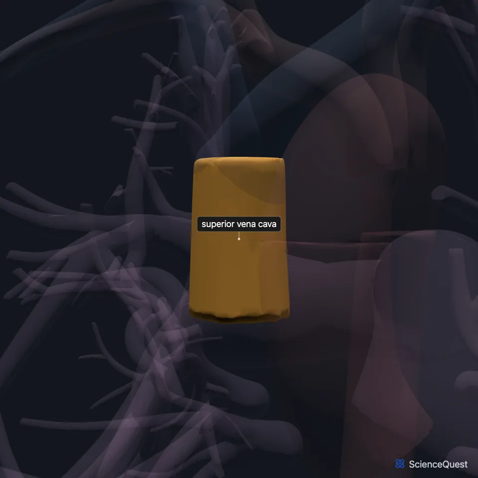
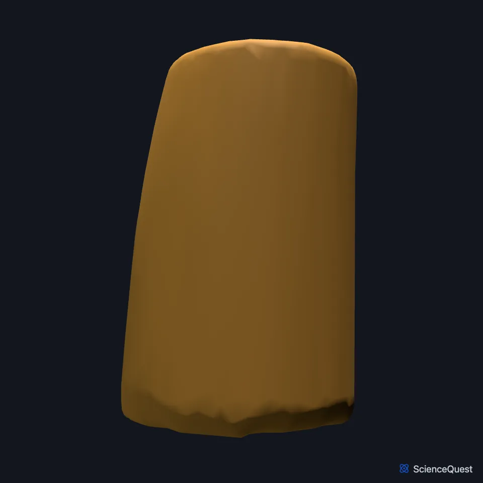
Where is the superior vena cava?
The right and left brachiocephalic veins join to form it behind the lower border of the first right costal cartilage. It runs straight down behind the second and third intercostal spaces. (StatPearls, Thorax, Superior Vena Cava)
It enters the right atrium at the level of the third costal cartilage, through an opening with no valve, at the upper and back part of the atrium. (StatPearls, Thorax, Superior Vena Cava; OpenStax A&P 2e, 19.1)
What does the superior vena cava drain?
It drains the head, neck, upper limbs and thorax. (OpenStax A&P 2e, 19.1; Gray’s Anatomy (1918), The Right Atrium; StatPearls, Thorax, Superior Vena Cava)
It also receives the azygos vein, which arches over the root of the right lung to join it at the level of the right second costal cartilage. (StatPearls, Thorax, Superior Vena Cava)
Each brachiocephalic vein forms where the subclavian vein meets the internal jugular vein, so the arms drain through the subclavians and the head through the internal jugulars. (StatPearls, Thorax, Brachiocephalic (Innominate) Veins)
What lies next to the superior vena cava?
The right phrenic nerve runs down its outer side, the right vagus nerve passes behind and to its inner side with the trachea, and the root of the right lung lies behind it. (StatPearls, Thorax, Superior Vena Cava)
Its lower part pierces the fibrous pericardium at the level of the azygos vein and stays inside it to its end. (StatPearls, Thorax, Superior Vena Cava)
How big is the superior vena cava?
In adults it is about 7 cm long and 2 cm wide. In children its size tracks height, and it reaches adult size at about 10 to 12 years. (StatPearls, Thorax, Superior Vena Cava)
What is a persistent left superior vena cava?
In about 0.3% of people an embryonic vein on the left persists as a left superior vena cava. When a normal right one is present too, the anomaly is a double superior vena cava. (StatPearls, Thorax, Superior Vena Cava)
Where the sources differ
The sources place the azygos vein’s entry by different landmarks: OpenStax at about the level of T2, StatPearls at the right second costal cartilage. (OpenStax A&P 2e, 20.5; StatPearls, Thorax, Superior Vena Cava)
| Feature | Detail and source |
|---|---|
| Formed by | Right and left brachiocephalic veins (StatPearls, Thorax, Superior Vena Cava) |
| Drains into | Right atrium, at the third costal cartilage (StatPearls, Thorax, Superior Vena Cava) |
| Size in adults | About 7 cm long, 2 cm wide (StatPearls, Thorax, Superior Vena Cava) |
Touches in this model
Measured on the BodyParts3D meshes: surfaces within 2 mm of the superior vena cava, nearest first.
- Arch of aorta in contact
- Ascending aorta in contact
- Left brachiocephalic vein in contact
- Right brachiocephalic vein in contact
- Right lobe of thymus in contact
- Wall of heart in contact
- Pulmonary artery 0.4 mm away
- Upper lobe of right lung 0.4 mm away
Sources
- StatPearls, Anatomy, Thorax, Superior Vena Cava. Hunter J. White; Michael P. Soos. Last update September 4, 2023. CC BY-NC-ND 4.0. Read 2026-10-25.
- OpenStax Anatomy and Physiology 2e, section 19.1, Heart Anatomy. CC BY-NC-SA 4.0. Read 2026-10-25.
- Anatomy of the Human Body (Gray’s Anatomy), 20th US edition, 1918, Angiology, The Heart, The Right Atrium, openings of the superior and inferior venae cavae; Internet Archive scan of the 1918 Lea and Febiger printing, terminology of 1918. Henry Gray; edited by Warren H. Lewis. Public domain. Read 2026-10-25.
- StatPearls, Anatomy, Thorax, Brachiocephalic (Innominate) Veins. Bruno Bordoni; Adekunle E. Omole. Last update December 9, 2025. CC BY-NC-ND 4.0. Read 2026-10-25.
- OpenStax Anatomy and Physiology 2e, section 20.5, Circulatory Pathways. CC BY-NC-SA 4.0. Read 2026-10-25.
The values are facts and free to use; this page’s selection and presentation are © 2026 ScienceQuest.