Thymus
The thymus is a two-lobed primary lymphoid organ in the superior mediastinum, behind the sternum. T cells made in the bone marrow travel to it to mature. (StatPearls, Head and Neck, Thymus; OpenStax A&P 2e, 21.1)
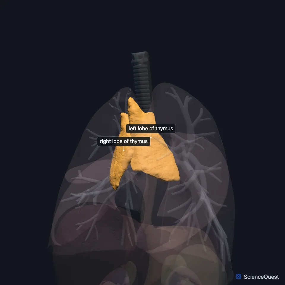
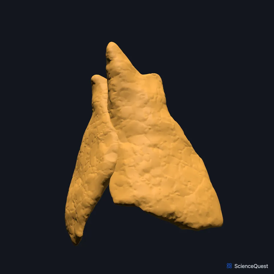
Where is the thymus?
Most of it lies in the anterior and anterosuperior mediastinum. Its two lobes meet near the manubrium of the sternum, and extensions can rise into the neck as far as the lower border of the thyroid gland. (StatPearls, Head and Neck, Thymus)
It usually measures 30 to 40 mm long and 25 to 35 mm wide, but its shape varies a great deal from person to person. (StatPearls, Head and Neck, Thymus)
What does the thymus do?
It is a primary lymphoid organ, where developing T cells, the thymocytes, mature and pass through selection. Cells whose receptors cannot recognize self-antigens die first, then cells that bind the body’s own antigens too strongly, and only cells that pass both tests leave to fight pathogens. (OpenStax A&P 2e, 21.1; StatPearls, Head and Neck, Thymus; Simonova et al. 2025, Int J Mol Sci)
Developing T cells pass through positive and then negative selection, and only those that pass both leave the thymus. Negative selection removes cells that bind the body’s own antigens strongly, which guards against autoimmunity. (StatPearls, Head and Neck, Thymus)
It is also an endocrine gland. Its epithelial cells make thymic hormones such as thymosin alpha-1, which act on T lymphocytes. (OpenStax A&P 2e, 17.10; Simonova et al. 2025, Int J Mol Sci)
What is the thymus made of?
A capsule encloses the gland, and septa of connective tissue divide each lobe into small lobules. Each lobule has an outer cortex crowded with immature T cells (thymocytes) and an inner medulla that holds fewer of them. (OpenStax A&P 2e, 21.1; Gray 1918, 4c. The Thymus; StatPearls, Head and Neck, Thymus; Simonova et al. 2025, Int J Mol Sci)
Hassall corpuscles (thymic corpuscles), found only in the thymus, help thymocytes mature and clear away cells that have died by apoptosis. (StatPearls, Head and Neck, Thymus)
How does the thymus change with age?
It is largest early in life and, from puberty, shrinks as fat replaces its tissue. The loss runs at about 3% a year until age 35 to 45, then about 1% a year. (StatPearls, Head and Neck, Thymus; OpenStax A&P 2e, 21.1)
The commonest variant is an extension up to the thyroid gland, seen in about a third of children who have a routine ultrasound of the neck. (StatPearls, Head and Neck, Thymus)
What lies next to the thymus?
In front are the manubrium, the deep cervical fascia and the sternohyoid and sternothyroid muscles. Behind are the great vessels of the superior mediastinum, the trachea and the front of the fibrous pericardium. (StatPearls, Head and Neck, Thymus)
At its lower end the right lobe lies between the right lung and the ascending aorta. (StatPearls, Head and Neck, Thymus)
Its arteries vary, but most often come from the inferior thyroid, internal thoracic, pericardiacophrenic or anterior intercostal arteries. Its veins drain mostly to the left brachiocephalic and internal thoracic veins. (StatPearls, Head and Neck, Thymus)
Its sympathetic nerves come from the superior cervical and stellate ganglia. Current anatomical evidence does not support a direct parasympathetic supply. (StatPearls, Head and Neck, Thymus)
| Feature | Detail and source |
|---|---|
| Region | Superior mediastinum, behind the manubrium (StatPearls, Head and Neck, Thymus) |
| Usual size | 30 to 40 mm long, 25 to 35 mm wide (StatPearls, Head and Neck, Thymus) |
| Develops from | Ventral third pharyngeal pouch (StatPearls, Head and Neck, Thymus) |
| Hormones | Thymic hormones, such as thymosin alpha-1 (OpenStax A&P 2e, 17.10; Simonova et al. 2025, Int J Mol Sci) |
Touches in this model
Measured on the BodyParts3D meshes: surfaces within 2 mm of the thymus, nearest first, 12 of 27.
- Bronchus in contact
- Upper lobe of left lung in contact
- Anterior interventricular branch of left coronary artery in contact
- Arch of aorta in contact
- Ascending aorta in contact
- Great cardiac vein in contact
- Left brachiocephalic vein in contact
- Left clavicle in contact
- Left sternothyroid in contact
- Left transversus thoracis in contact
- Manubrium in contact
- Marginal branch of right coronary artery in contact
Sources
- StatPearls, Anatomy, Head and Neck, Thymus. Kailey Remien; Felix Jozsa; Arif Jan. Last update June 23, 2025. CC BY-NC-ND 4.0. Read 2026-10-25.
- OpenStax Anatomy and Physiology 2e, section 21.1, Anatomy of the Lymphatic and Immune Systems. CC BY-NC-SA 4.0. Read 2026-10-25.
- International Journal of Molecular Sciences, Aging and Thymosin Alpha-1, Int J Mol Sci 26(23), article 11470 (PMC12692621). Simonova MA, Ivanov I, Shoshina NS, Komyakova AM, Makarov DA, et al.. CC BY 4.0. Read 2026-10-25.
- OpenStax Anatomy and Physiology 2e, section 17.10, Organs with Secondary Endocrine Functions. CC BY-NC-SA 4.0. Read 2026-10-25.
- Gray’s Anatomy of the Human Body, 20th US edition (1918), XI. Splanchnology, 4c. The Thymus (1918 terms: follicles for the small lobules, concentric corpuscles of Hassall for thymic corpuscles). Henry Gray. Public domain. Read 2026-10-25.
The values are facts and free to use; this page’s selection and presentation are © 2026 ScienceQuest.