Marginal branch of right coronary artery
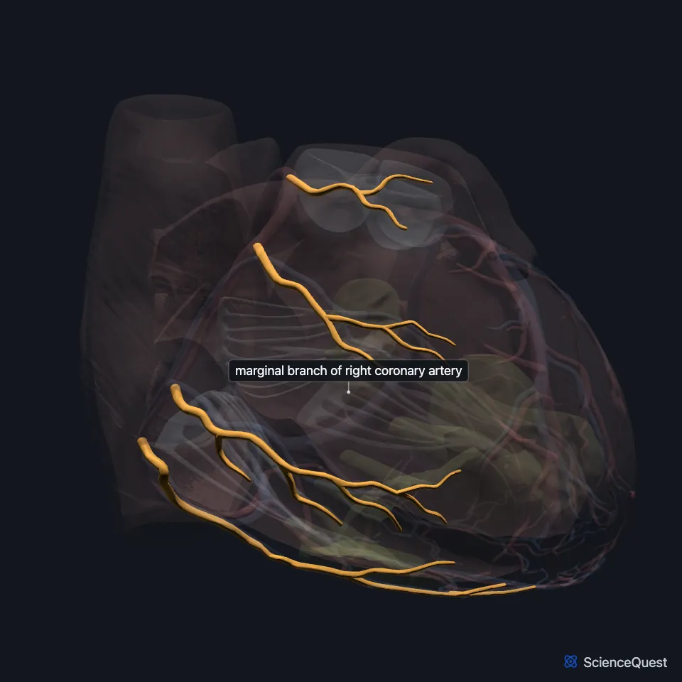
The right marginal artery is a branch of the right coronary artery. It runs along the acute margin of the heart, the lower border of its front surface. (StatPearls, Heart Arteries)
Where is the right marginal artery?
It leaves the right coronary artery as that artery runs along the coronary sulcus, below the right atrium. (StatPearls, Heart Arteries; OpenStax A&P 2e, 19.1)
What does the right marginal artery supply?
It supplies the right ventricle, the superficial parts of its wall. (OpenStax A&P 2e, 19.1; StatPearls, Heart Arteries)
What runs with the right marginal artery?
The small cardiac vein runs beside it on the front of the heart. That vein collects blood from the back of the right chambers and empties into the coronary sinus. (StatPearls, Heart Veins)
The right marginal vein lies on the lowest margin of the front of the heart, the same border the artery follows. It drains the front and diaphragmatic walls of the right ventricle and joins the small cardiac vein in the coronary sulcus. (StatPearls, Heart Veins and Heart Arteries)
Is there only one right marginal artery?
The right coronary artery gives off the right marginal artery as it runs along the coronary sulcus. (OpenStax A&P 2e, 19.1; StatPearls, Heart Arteries; Gray 1918, 2. The Aorta)
| Feature | Detail and source |
|---|---|
| Branch of | Right coronary artery (StatPearls, Heart Arteries) |
| Runs along | The acute margin, the anteroinferior border of the heart (StatPearls, Heart Arteries) |
| Supplies | The right ventricle, on both its surfaces (OpenStax A&P 2e, 19.1; Gray 1918, 2. The Aorta; StatPearls, Heart Arteries) |
Touches in this model
Measured on the BodyParts3D meshes: surfaces within 2 mm of the marginal branch of right coronary artery, nearest first, 12 of 14.
- Anterior papillary muscle of right ventricle in contact
- Middle cardiac vein in contact
- Set of anterior cardiac veins in contact
- Trunk of right coronary artery in contact
- Wall of heart in contact
- Left lobe of thymus in contact
- Middle lobe of lung in contact
- Right lobe of thymus in contact
- Upper lobe of left lung in contact
- Posterior interventricular branch of right coronary artery 0.2 mm away
- Diaphragm 0.3 mm away
- Posterior papillary muscle of right ventricle 0.7 mm away
Sources
- StatPearls, Anatomy, Thorax, Heart Arteries. Raheel Chaudhry; Sajedur Rahman; Mark A. Law. Last update July 24, 2023. CC BY-NC-ND 4.0. Read 2026-10-25.
- OpenStax Anatomy and Physiology 2e, section 19.1, Heart Anatomy. CC BY-NC-SA 4.0. Read 2026-10-25.
- StatPearls, Anatomy, Thorax, Heart Veins. Julia H. Miao; Amgad N. Makaryus. Last update July 24, 2023. CC BY-NC-ND 4.0. Read 2026-10-25.
- Gray’s Anatomy of the Human Body, 20th US edition (1918), VI. The Arteries, 2. The Aorta (1918 terms: longitudinal sulcus for interventricular sulcus, anterior aortic sinuses for the coronary sinuses of the aorta). Henry Gray. Public domain. Read 2026-10-25.
The values are facts and free to use; this page’s selection and presentation are © 2026 ScienceQuest.