Bronchus
The bronchi are the airways that continue the trachea into the lungs. With the trachea they form the tracheobronchial tree, part of the conducting zone of the respiratory system. (StatPearls, Bronchial)
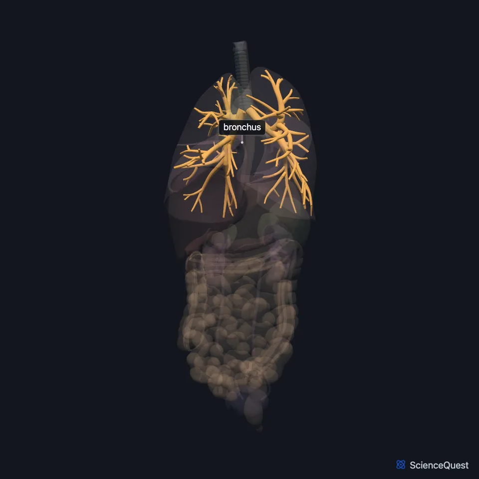
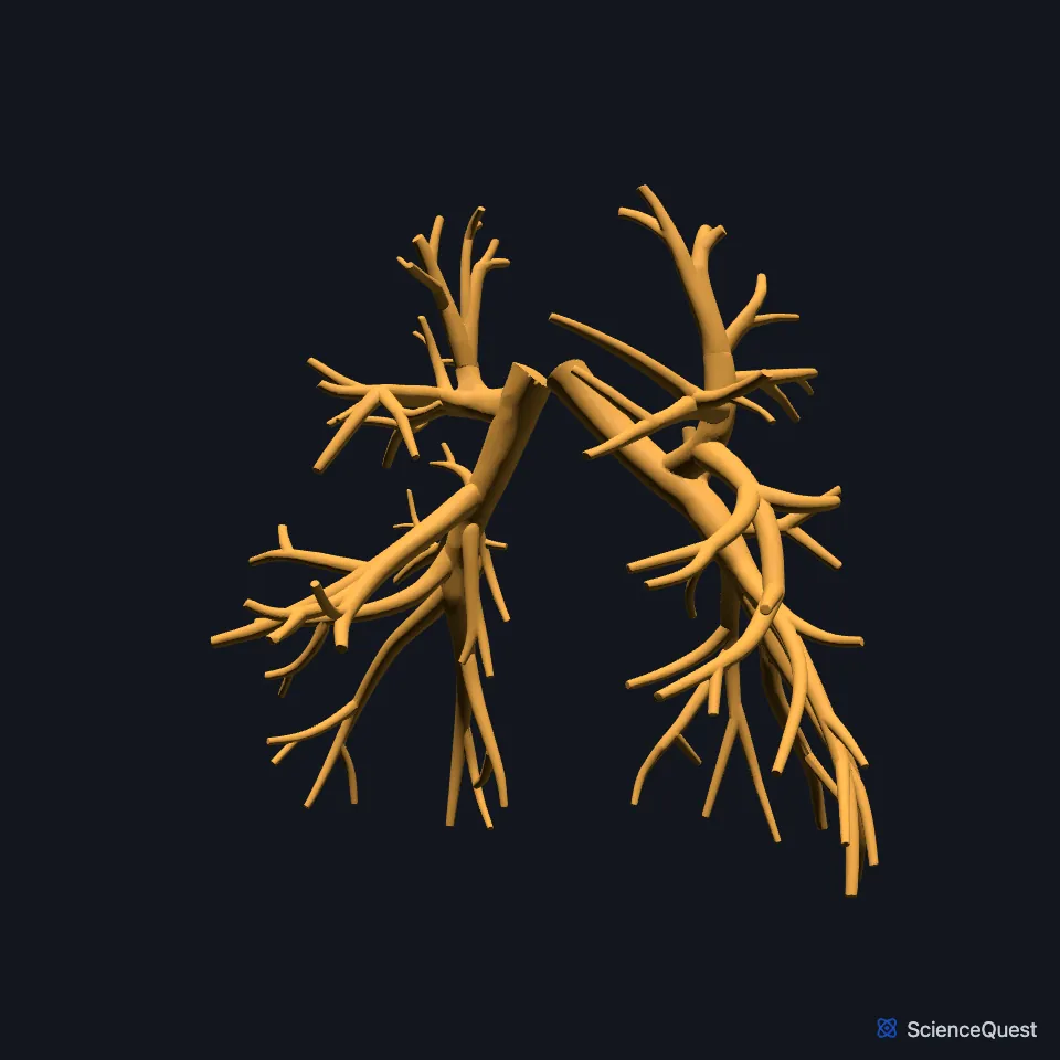
Where are the bronchi?
The trachea divides into the right and left main bronchi at the carina, a ridge of cartilage at its lower end. The main bronchi enter the lungs at the hilum, together with the pulmonary vessels, bronchial vessels and lymphatics. (OpenStax A&P 2e, 22.1; StatPearls, Tracheobronchial Tree, Bronchial and Thorax, Lungs)
The right main bronchus is wider, shorter and more vertical than the left. The left one runs more horizontally, passing below the arch of the aorta and in front of the esophagus and thoracic aorta to reach the left lung. (StatPearls, Bronchial)
How do the bronchi branch?
Each main bronchus divides into lobar bronchi, one per lobe: three on the right and two on the left. These split into segmental bronchi, and each segmental bronchus supplies one bronchopulmonary segment. (StatPearls, Bronchial)
Beyond the segmental bronchi the airways become bronchioles. The terminal bronchioles end the conducting zone, and respiratory bronchioles lead on to the alveolar ducts and sacs, where gas exchange takes place. (StatPearls, Bronchial)
Down the tree, cartilage thins out and smooth muscle increases. The bronchioles have no cartilage and keep their shape with smooth muscle and elastic fibers. (StatPearls, Bronchial)
Like the trachea, the bronchi are lined by pseudostratified ciliated columnar epithelium with mucus-producing goblet cells. (OpenStax A&P 2e, 22.1; StatPearls, Bronchial)
What do the bronchi do?
They carry air into and out of each lung, and their mucous lining traps foreign material you breathe in. (OpenStax A&P 2e, 22.1; StatPearls, Bronchial)
Smooth muscle in the wall narrows or widens the airway to control airflow. Parasympathetic fibers from the vagus contract it, causing bronchoconstriction, and sympathetic fibers relax it, causing bronchodilation. (StatPearls, Bronchial; OpenStax A&P 2e, 22.2)
What supplies the bronchi, and what runs with them?
Bronchial arteries, usually from the start of the descending aorta, supply the lower trachea, the carina and the bronchi. (StatPearls, Tracheobronchial Tree)
The right bronchial vein drains into the azygos vein. The left drains into the accessory hemiazygos vein or the left superior intercostal vein. (StatPearls, Bronchial)
Branches of the pulmonary artery run alongside the bronchial tree and carry deoxygenated blood out to the alveolar capillaries. (OpenStax A&P 2e, 22.2; StatPearls, Airway)
Lymph from the lung passes from the hilum to the tracheobronchial nodes at the tracheal bifurcation, then to the paratracheal nodes. (StatPearls, Bronchial)
Why do inhaled objects tend to go right?
Inhaled foreign bodies lodge in the right lung more often than the left, because the right main bronchus is wider, shorter and more vertical. (StatPearls, Bronchial)
Where the sources differ
The sources describe the cartilage of the bronchi differently. OpenStax speaks of rings of cartilage like those of the trachea; StatPearls gives the trachea C-shaped cartilage and the bronchi plates of hyaline cartilage. (OpenStax A&P 2e, 22.1; StatPearls, Bronchial)
They also differ on the left lung’s bronchopulmonary segments, each supplied by its own segmental bronchus: 8 to 10 in the StatPearls bronchi article, 8 to 9 in the StatPearls lungs article. (StatPearls, Bronchial and Thorax, Lungs)
| Feature | Detail and source |
|---|---|
| Main bronchi | Two, from the carina; the right is wider, shorter and more vertical (StatPearls, Bronchial) |
| Lobar bronchi | One per lobe: three right, two left (StatPearls, Bronchial) |
| Lining | Pseudostratified ciliated columnar epithelium with goblet cells (OpenStax A&P 2e, 22.1; StatPearls, Bronchial) |
| Arteries | Bronchial arteries, usually from the proximal descending aorta (StatPearls, Tracheobronchial Tree) |
| Nerves | Pulmonary plexus: vagal fibers constrict, sympathetic fibers dilate (StatPearls, Bronchial) |
Touches in this model
Measured on the BodyParts3D meshes: surfaces within 2 mm of the bronchus, nearest first, 12 of 28.
- Left lobe of thymus in contact
- Lower lobe of left lung in contact
- Lower lobe of right lung in contact
- Middle lobe of lung in contact
- Right lobe of thymus in contact
- Trachea in contact
- Upper lobe of left lung in contact
- Upper lobe of right lung in contact
- Diaphragm in contact
- Innermost intercostal muscle in contact
- Left subclavian artery in contact
- Left transversus abdominis in contact
Sources
- StatPearls, Anatomy, Thorax, Bronchial. Christina Amador; Carly Weber; Matthew A. Varacallo. Last update August 8, 2023. CC BY-NC-ND 4.0. Read 2026-10-25.
- OpenStax Anatomy and Physiology 2e, section 22.1, Organs and Structures of the Respiratory System. CC BY-NC-SA 4.0. Read 2026-10-25.
- StatPearls, Anatomy, Thorax, Tracheobronchial Tree. Ryan P. Downey; Navdeep S. Samra. Last update July 24, 2023. CC BY-NC-ND 4.0. Read 2026-10-25.
- StatPearls, Anatomy, Thorax, Lungs. Raheel Chaudhry; Adekunle E. Omole; Bruno Bordoni. Last update April 20, 2024. CC BY-NC-ND 4.0. Read 2026-10-25.
- OpenStax Anatomy and Physiology 2e, section 22.2, The Lungs. CC BY-NC-SA 4.0. Read 2026-10-25.
- StatPearls, Anatomy, Airway. Steffi C. Nata; Marjorie V. Launico. Last update September 15, 2025. CC BY-NC-ND 4.0. Read 2026-10-25.
The values are facts and free to use; this page’s selection and presentation are © 2026 ScienceQuest.