Transversus abdominis
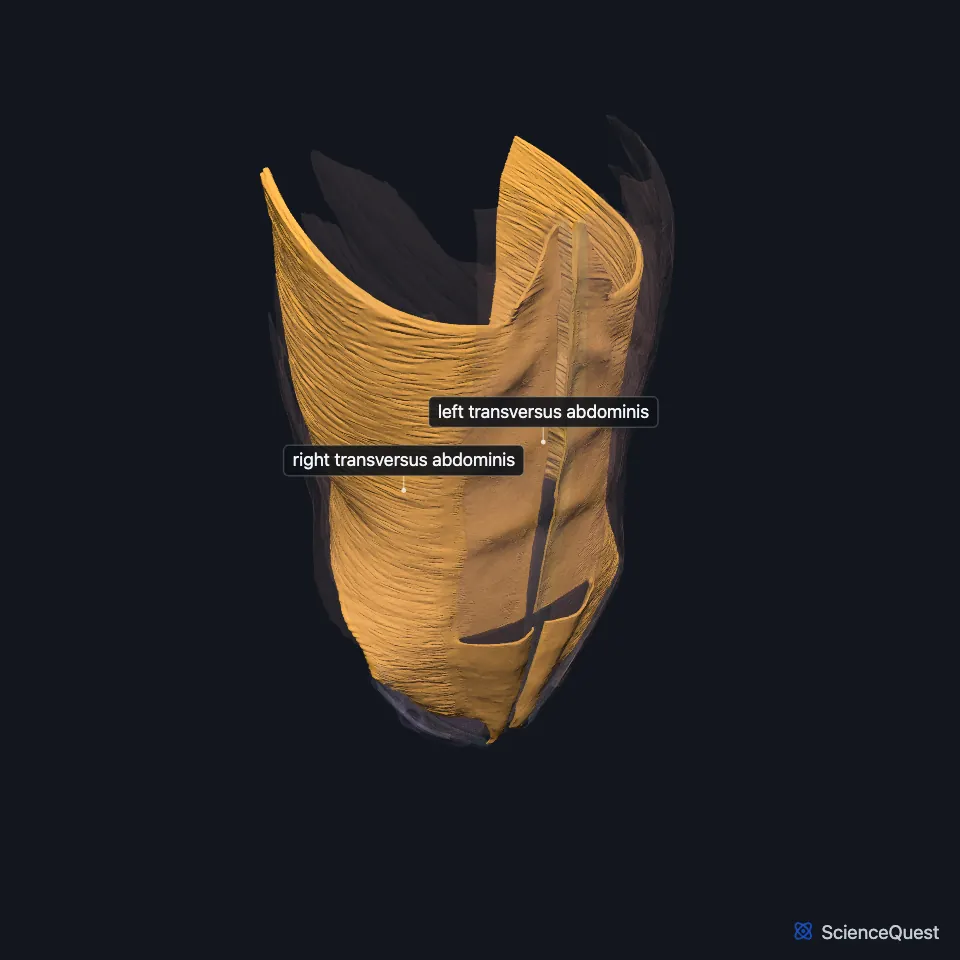
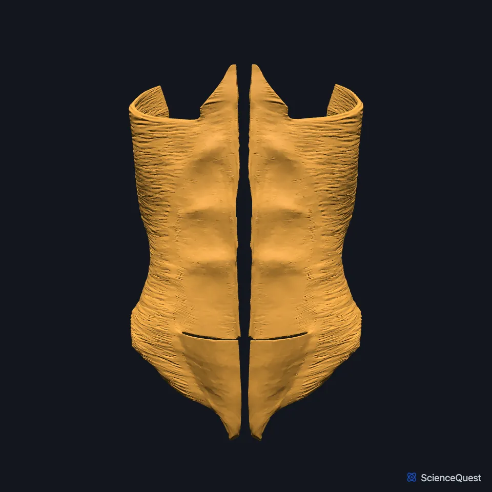
The transversus abdominis is the thinnest and deepest of the three flat muscles of the abdominal wall. Its fibers run across the abdomen, like the front of a belt. (StatPearls, Anterolateral Abdominal Wall; OpenStax A&P 2e, 11.4)
Open it in the Abdominal Muscles Explorer See it alone in 3D
Where is the transversus abdominis?
It lies under the internal oblique. Beneath it lies the transversalis fascia, and beneath that the parietal peritoneum. (OpenStax A&P 2e, 11.4; StatPearls, Anterolateral Abdominal Wall Muscles and Anterolateral Abdominal Wall)
StatPearls’ quadratus lumborum article describes the quadratus lumborum as a backward continuation of the transversus abdominis, lying nearer the midline than its aponeurosis. (StatPearls, Quadratus Lumborum)
What does the transversus abdominis attach to?
Both StatPearls accounts have it arising from costal cartilages, the lumbar region and the iliac crest. Its fibers run across and end in an aponeurosis that enters the rectus sheath and reaches the linea alba. (StatPearls, Abdominal Wall, Anterolateral Abdominal Wall and Anterolateral Abdominal Wall Muscles)
Above the arcuate line its aponeurosis passes behind the rectus abdominis and forms part of the back wall of the sheath. Below the line it passes in front. (StatPearls, Abdominal Wall)
Its lower edge and the internal oblique’s form the conjoint tendon. Its costal origin interlocks with the costal part of the diaphragm. (StatPearls, Abdominal Wall and Thorax: Diaphragm)
What does the transversus abdominis do?
It compresses the abdominal contents. OpenStax lists the squeeze it gives in forceful breathing out, defecation, urination and childbirth. (StatPearls, Abdominal Wall; OpenStax A&P 2e, 11.4)
StatPearls leaves it out of the accessory breathing muscles that pull the ribs down in active expiration, a role it gives the other anterolateral muscles. (StatPearls, Abdominal Wall)
Which nerves run on the transversus abdominis?
The nerves of the abdominal wall, from the anterior rami of T7 to T12, run between it and the internal oblique. The iliohypogastric and ilioinguinal nerves supply its lowest fibers. (StatPearls, Anterolateral Abdominal Wall and Abdominal Wall)
Because so many nerves share that plane, local anesthetic placed there numbs a wide field of the wall; this is the transversus abdominis plane (TAP) block. (StatPearls, Abdominal Wall)
Does the transversus abdominis reach into the rectus sheath?
One study found its muscle fibers inside the back wall of the rectus sheath, mostly high up near the costal margin and fewer lower down. StatPearls adds that the textbook pattern of the anterolateral wall appears in just over half of the bodies examined. (StatPearls, Rectus Sheath and Anterolateral Abdominal Wall)
Where the sources differ
The StatPearls abdominal wall article starts its costal origin at the fifth to tenth costal cartilages and adds the lateral inguinal ligament. The anterolateral abdominal wall article gives the lower six costal cartilages and adds the lumbar vertebrae and the iliopsoas fascia. (StatPearls, Abdominal Wall and Anterolateral Abdominal Wall)
OpenStax’s table gives its origin as the ilium and ribs 5 to 10 and its insertion as the sternum, the linea alba and the pubis. StatPearls inserts it on the rectus sheath and the linea alba, through its aponeurosis. (OpenStax A&P 2e, 11.4; StatPearls, Abdominal Wall and Anterolateral Abdominal Wall Muscles)
| Feature | Detail and source |
|---|---|
| Origin | Costal cartilages (the fifth to tenth, or the lower six), lumbar fascia or vertebrae, iliac crest (StatPearls, Abdominal Wall and Anterolateral Abdominal Wall) |
| Insertion | Rectus sheath and linea alba, through its aponeurosis (StatPearls, Abdominal Wall and Anterolateral Abdominal Wall Muscles) |
| Action | Compresses the abdominal contents (StatPearls, Abdominal Wall) |
| Nerve | Anterior rami of T7 to T12; iliohypogastric and ilioinguinal nerves to its lowest fibers (StatPearls, Anterolateral Abdominal Wall and Abdominal Wall) |
Touches in this model
Measured on the BodyParts3D meshes: surfaces within 2 mm of the transversus abdominis, nearest first, 12 of 69.
- Left inguinal ligament in contact
- Left internal oblique in contact
- Left pyramidalis in contact
- Left quadratus lumborum in contact
- Left rectus abdominis in contact
- Linea alba in contact
- Right inguinal ligament in contact
- Right internal oblique in contact
- Right pyramidalis in contact
- Right quadratus lumborum in contact
- Right rectus abdominis in contact
- Bronchus in contact
Sources
- StatPearls, Anatomy, Abdomen and Pelvis: Anterolateral Abdominal Wall. Kevin Seeras; Ryan N. Qasawa; Ricky Ju; Shivana Prakash. Last update July 24, 2023. CC BY-NC-ND 4.0. Read 2026-10-25.
- OpenStax Anatomy and Physiology 2e, section 11.4, Axial Muscles of the Abdominal Wall, and Thorax. CC BY-NC-SA 4.0. Read 2026-10-25.
- StatPearls, Anatomy, Anterolateral Abdominal Wall Muscles. Matthew A. Varacallo; Stephanie Scharbach; Mohammed A. Al-Dhahir. Last update July 24, 2023. CC BY-NC-ND 4.0. Read 2026-10-25.
- StatPearls, Anatomy, Abdomen and Pelvis, Quadratus Lumborum. Bruno Bordoni; Reddog E. Sina; Matthew A. Varacallo. Last update July 17, 2024. CC BY-NC-ND 4.0. Read 2026-10-25.
- StatPearls, Anatomy, Abdomen and Pelvis: Abdominal Wall. William Flynn; Paula Vickerton. Last update July 24, 2023. CC BY-NC-ND 4.0. Read 2026-10-25.
- StatPearls, Anatomy, Thorax: Diaphragm. Kanwal Naveen S. Bains; Sarang Kashyap; Sarah L. Lappin. Last update July 24, 2023. CC BY-NC-ND 4.0. Read 2026-10-25.
- StatPearls, Anatomy, Abdomen and Pelvis, Rectus Sheath. Karlin E. Sevensma; Logan Leavitt; Kerent D. Pihl. Last update July 24, 2023. CC BY-NC-ND 4.0. Read 2026-10-25.
The values are facts and free to use; this page’s selection and presentation are © 2026 ScienceQuest.