Inferior vena cava
The inferior vena cava is the large vein that returns blood from the lower limbs and abdomen to the right atrium. It has the widest diameter of any vein. (StatPearls, Abdomen and Pelvis: Inferior Vena Cava)
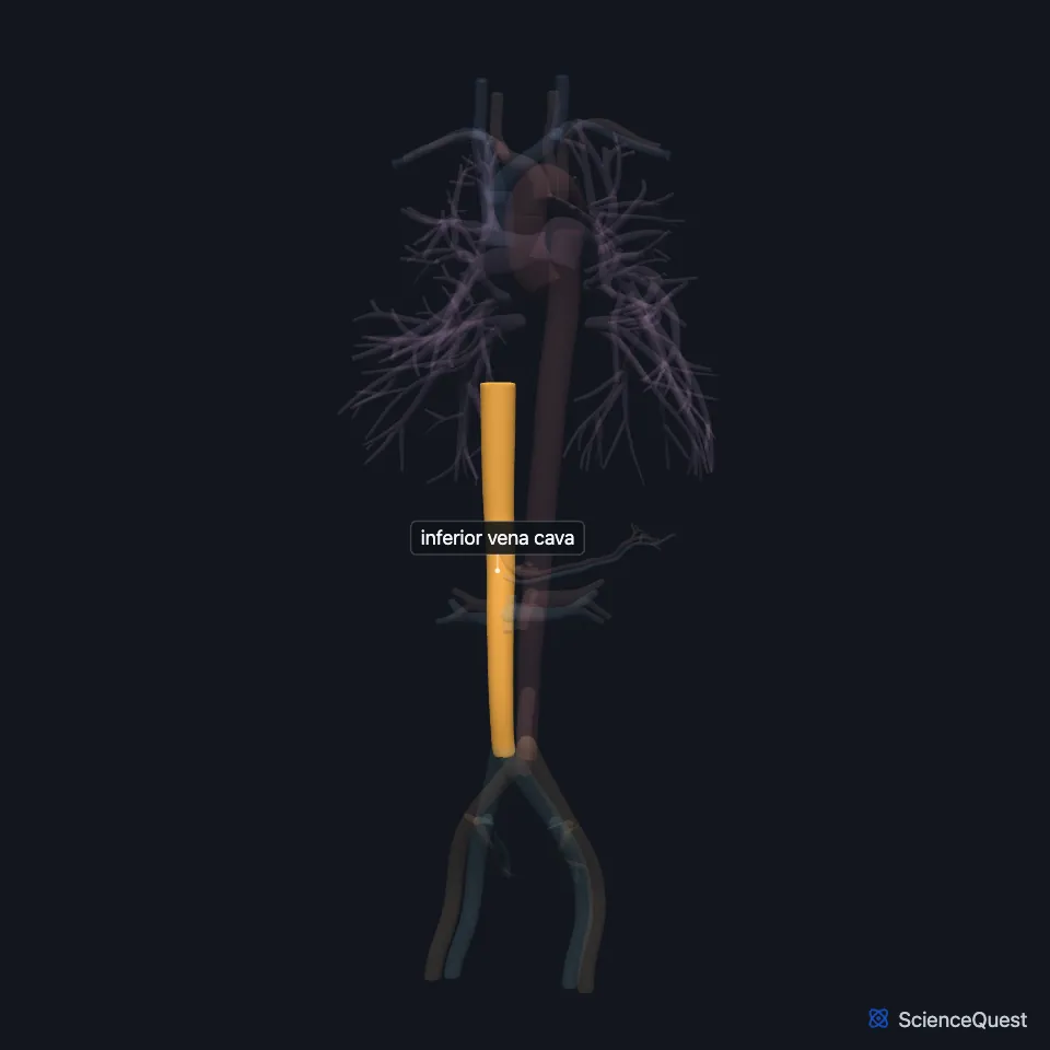
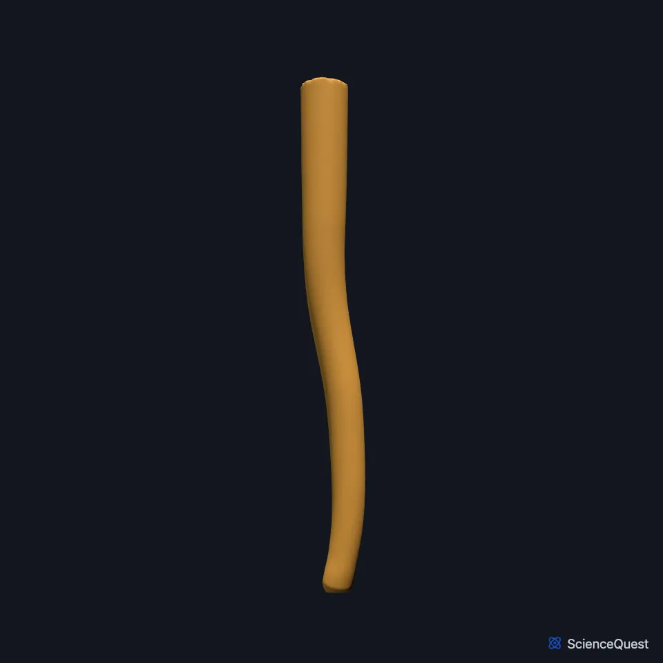
Where is the inferior vena cava?
The right and left common iliac veins join to form it, usually at the level of L5. It runs up the right front side of the vertebral column, behind the peritoneum. (StatPearls, Abdomen and Pelvis: Inferior Vena Cava)
It passes through the central tendon of the diaphragm at about T8 and enters the back of the right atrium, below the opening of the superior vena cava. (StatPearls, Abdomen and Pelvis: Inferior Vena Cava)
What does the inferior vena cava drain?
It returns blood from everything below the diaphragm: the lower limbs, pelvis and abdomen. (OpenStax A&P 2e, 20.5; Gray’s Anatomy (1918), The Right Atrium; StatPearls, Abdomen and Pelvis: Inferior Vena Cava and Thorax, Superior Vena Cava)
Unlike many veins, it has no one-way valves. Breathing drives the flow: as the diaphragm contracts, the drop in chest pressure draws blood from the abdominal part of the vein into the thoracic part and the heart. (StatPearls, Abdomen and Pelvis: Inferior Vena Cava)
Blood from the gut passes through the liver first and reaches the inferior vena cava through the hepatic veins. (StatPearls, Abdomen and Pelvis: Inferior Vena Cava)
Which veins join the inferior vena cava?
Typical levels of entry: hepatic and inferior phrenic veins at T8, the right suprarenal and renal veins at L1, the right gonadal vein at L2, lumbar veins from L1 to L5 and the common iliac veins at L5. (StatPearls, Abdomen and Pelvis: Inferior Vena Cava)
Each kidney drains through its renal vein into it, at about the level of L1. (OpenStax A&P 2e, 20.5; StatPearls, Abdomen and Pelvis, Renal Veins and Abdomen and Pelvis: Inferior Vena Cava)
Why are the left side veins longer?
The inferior vena cava lies to the right of the vertebral column, so veins that reach it from the left, such as the left renal vein, are longer than their right-hand partners. StatPearls adds that the left adrenal and gonadal veins join the left renal vein before reaching it. (OpenStax A&P 2e, 20.5; StatPearls, Abdomen and Pelvis: Inferior Vena Cava and Abdomen and Pelvis, Renal Veins)
On the left, the adrenal and gonadal veins drain into the left renal vein first. On the right they open into the inferior vena cava itself. (StatPearls, Abdomen and Pelvis: Inferior Vena Cava)
Which collateral route links the two venae cavae?
Ascending lumbar veins link the lumbar veins to the azygos vein, a collateral route between the inferior and superior vena cava. (StatPearls, Abdomen and Pelvis: Inferior Vena Cava)
How does the inferior vena cava vary?
A left inferior vena cava occurs in an estimated 0.2% to 0.5% of people, and a double inferior vena cava in 0.2% to 0.3%. (StatPearls, Abdomen and Pelvis: Inferior Vena Cava)
| Feature | Detail and source |
|---|---|
| Formed by | Right and left common iliac veins, usually at L5 (StatPearls, Abdomen and Pelvis: Inferior Vena Cava) |
| Passes the diaphragm | Through the central tendon, about T8 (StatPearls, Abdomen and Pelvis: Inferior Vena Cava) |
| Drains into | Right atrium, below the superior vena cava’s opening (OpenStax A&P 2e, 19.1; StatPearls, Thorax, Superior Vena Cava; Gray’s Anatomy (1918), The Right Atrium) |
| Valves | No one-way venous valves (StatPearls, Abdomen and Pelvis: Inferior Vena Cava) |
Touches in this model
Measured on the BodyParts3D meshes: surfaces within 2 mm of the inferior vena cava, nearest first, 12 of 14.
- Left common iliac vein in contact
- Left renal vein in contact
- Right common iliac artery in contact
- Right common iliac vein in contact
- Right renal artery in contact
- Right renal vein in contact
- Splenic vein in contact
- Superior mesenteric vein in contact
- Diaphragm in contact
- Liver in contact
- Pancreas in contact
- Wall of heart in contact
Sources
- StatPearls, Anatomy, Abdomen and Pelvis: Inferior Vena Cava. William D. Tucker; Ribesh Shrestha; Bracken Burns. Last update July 24, 2023. CC BY-NC-ND 4.0. Read 2026-10-25.
- OpenStax Anatomy and Physiology 2e, section 20.5, Circulatory Pathways. CC BY-NC-SA 4.0. Read 2026-10-25.
- Anatomy of the Human Body (Gray’s Anatomy), 20th US edition, 1918, Angiology, The Heart, The Right Atrium, openings of the superior and inferior venae cavae; Internet Archive scan of the 1918 Lea and Febiger printing, terminology of 1918. Henry Gray; edited by Warren H. Lewis. Public domain. Read 2026-10-25.
- StatPearls, Anatomy, Thorax, Superior Vena Cava. Hunter J. White; Michael P. Soos. Last update September 4, 2023. CC BY-NC-ND 4.0. Read 2026-10-25.
- StatPearls, Anatomy, Abdomen and Pelvis, Renal Veins. Cole S. Bowdino; Justin Owens; Palma M. Shaw. Last update January 2, 2023. CC BY-NC-ND 4.0. Read 2026-10-25.
- OpenStax Anatomy and Physiology 2e, section 19.1, Heart Anatomy. CC BY-NC-SA 4.0. Read 2026-10-25.
The values are facts and free to use; this page’s selection and presentation are © 2026 ScienceQuest.