Pulmonary valve
The pulmonary valve, also called the pulmonic valve or the pulmonary semilunar valve, sits where the pulmonary trunk leaves the right ventricle. It has three small flaps of the heart’s lining reinforced with fibrous connective tissue. (OpenStax A&P 2e, 19.1; FMA, FMA7246; Gray 1918, 4b. The Heart; StatPearls, Histology, Heart)
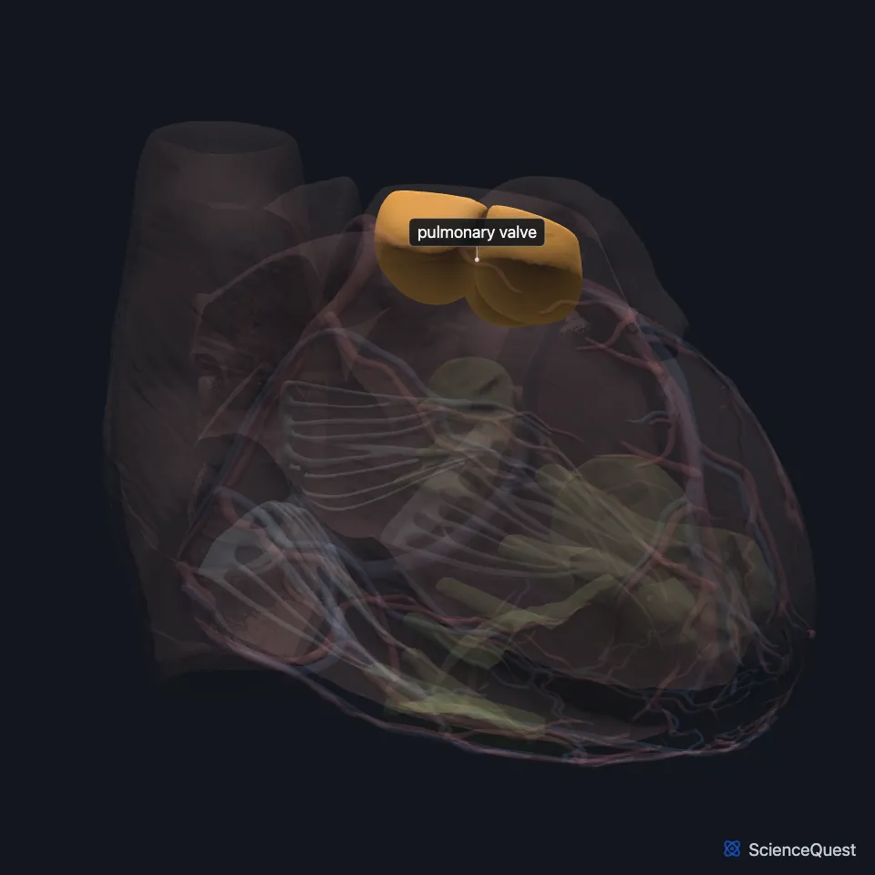
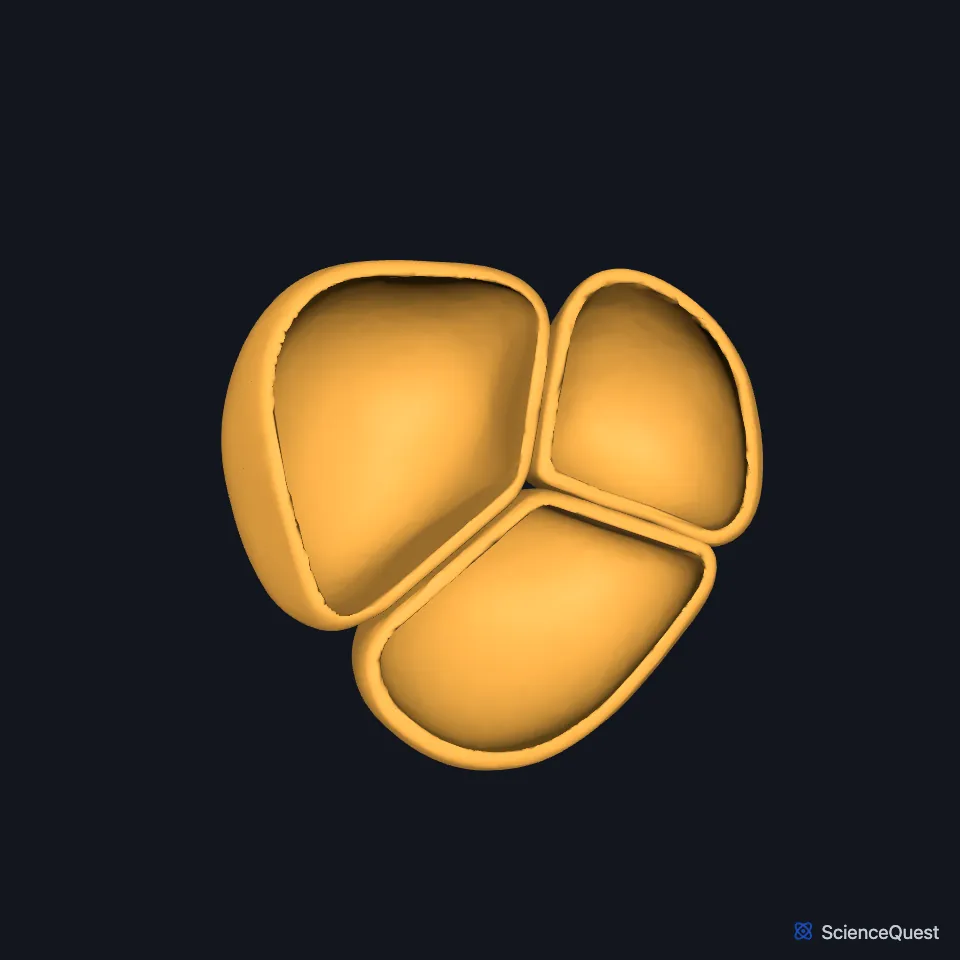
Where is the pulmonary valve?
It separates the right ventricle from the pulmonary trunk, where the top of the ventricle narrows toward the trunk. (StatPearls, Heart Pulmonic Valve; OpenStax A&P 2e, 19.1)
The muscle of the subpulmonary infundibulum lifts it above the ventricular septum, which makes it the highest of the heart valves. (StatPearls, Heart Pulmonic Valve)
Clinicians listen for its closure in the second left intercostal space. (StatPearls, Heart Sounds)
What are the parts of the pulmonary valve?
Its three cusps are the anterior, the left and the right, with a commissure between each pair. Below their free edges the cusps overlap in an area called the lunula, which helps seal the opening. (StatPearls, Heart Pulmonic Valve)
A small fibrous thickening at the center of the lunula, the nodule of Arantius, helps close the opening. (StatPearls, Heart Pulmonic Valve)
It has no papillary muscles or chordae tendineae. Its three cusps are folds of the heart’s lining strengthened by fibrous tissue, with a pouch behind each. (OpenStax A&P 2e, 19.1; StatPearls, Histology, Heart; Gray 1918, 4b. The Heart)
The valve is about 20 mm across. It has no blood supply or lymphatic drainage of its own. (StatPearls, Heart Pulmonic Valve)
What does the pulmonary valve do?
It opens when the right ventricle contracts and lets blood out to the lungs. It closes while the ventricle relaxes and refills. (StatPearls, Heart Pulmonic Valve)
As the ventricle relaxes, blood flowing back from the pulmonary trunk fills the pocket-like flaps. That closes the valve and makes an audible sound. (OpenStax A&P 2e, 19.1; Gray 1918, 4b. The Heart; StatPearls, Cardiac Cycle)
Its closure is the later, softer part of the second heart sound, S2, after the aortic valve’s. When you breathe in, the pulmonary closure comes later still, and S2 can be heard as two sounds. (StatPearls, Heart Sounds)
What lies around the pulmonary valve?
Above it, the pulmonary trunk divides into the left and right pulmonary arteries, which carry blood to each lung. (OpenStax A&P 2e, 19.1; Gray 1918, The Pulmonary Artery)
A muscular fold, the ventriculoinfundibular fold, separates it from the tricuspid valve. (StatPearls, Heart Pulmonic Valve)
Its annulus and the cardiac fibrous skeleton link it to the other heart valves and anchor all of them in the heart muscle. (StatPearls, Heart Pulmonic Valve)
What can go wrong with the pulmonary valve?
The valve can form with one or two cusps instead of three, or with no opening at all (atresia). Any of these abnormalities can cause congenital semilunar valve disease. (StatPearls, Heart Pulmonic Valve)
| Feature | Detail and source |
|---|---|
| Between | Right ventricle and pulmonary trunk (StatPearls, Heart Pulmonic Valve) |
| Cusps | Three: anterior, left and right (StatPearls, Heart Pulmonic Valve) |
| Other names | Pulmonic valve, pulmonary semilunar valve (OpenStax A&P 2e, 19.1; FMA, FMA7246; Gray 1918, 4b. The Heart) |
| Diameter | About 20 mm (StatPearls, Heart Pulmonic Valve) |
| Closes | As the ventricles relax, making part of the second heart sound (S2) (OpenStax A&P 2e, 19.3; StatPearls, Cardiac Cycle) |
Touches in this model
Measured on the BodyParts3D meshes: surfaces within 2 mm of the pulmonary valve, nearest first.
- Wall of heart in contact
- Pulmonary artery in contact
- Right lobe of thymus 0.6 mm away
- Ascending aorta 1.1 mm away
- Trunk of right coronary artery 1.3 mm away
- Left lobe of thymus 1.3 mm away
Sources
- OpenStax Anatomy and Physiology 2e, section 19.1, Heart Anatomy. CC BY-NC-SA 4.0. Read 2026-10-25.
- Foundational Model of Anatomy, via the EMBL-EBI Ontology Lookup Service, Pulmonary valve (FMA7246), preferred name and synonyms. Not printed on the page. Read 2026-10-25.
- Gray’s Anatomy of the Human Body, 20th US edition (1918), V. Angiology, 4b. The Heart (1918 terms: longitudinal sulci for interventricular sulci, musculi papillares for papillary muscles). Henry Gray. Public domain. Read 2026-10-25.
- StatPearls, Histology, Heart. Anita Arackal; Khalid Alsayouri. Last update January 2, 2023. CC BY-NC-ND 4.0. Read 2026-10-25.
- StatPearls, Anatomy, Thorax, Heart Pulmonic Valve. Joshua Henrina Sundjaja; Bruno Bordoni. Last update July 24, 2023. CC BY-NC-ND 4.0. Read 2026-10-25.
- StatPearls, Physiology, Heart Sounds. Sean Dornbush; Andre E. Turnquest. Last update July 17, 2023. CC BY-NC-ND 4.0. Read 2026-10-25.
- StatPearls, Physiology, Cardiac Cycle. Joshua D. Pollock; Amgad N. Makaryus. Last update October 3, 2022. CC BY-NC-ND 4.0. Read 2026-10-25.
- Gray’s Anatomy of the Human Body, 20th US edition (1918), VI. The Arteries, 1. Introduction (The Pulmonary Artery). Henry Gray. Public domain. Read 2026-10-25.
- OpenStax Anatomy and Physiology 2e, section 19.3, Cardiac Cycle. CC BY-NC-SA 4.0. Read 2026-10-25.
The values are facts and free to use; this page’s selection and presentation are © 2026 ScienceQuest.