Mitral valve
The mitral valve guards the opening between the left atrium and the left ventricle; its other names are the bicuspid valve and the left atrioventricular valve. It has two cusps, where the tricuspid valve has three. (OpenStax A&P 2e, 19.1; Gray 1918, 4b. The Heart; FMA, FMA7235)
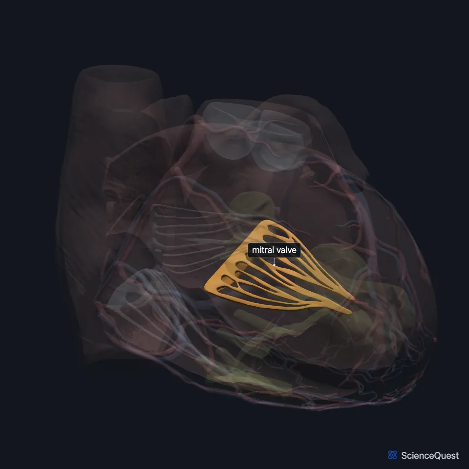
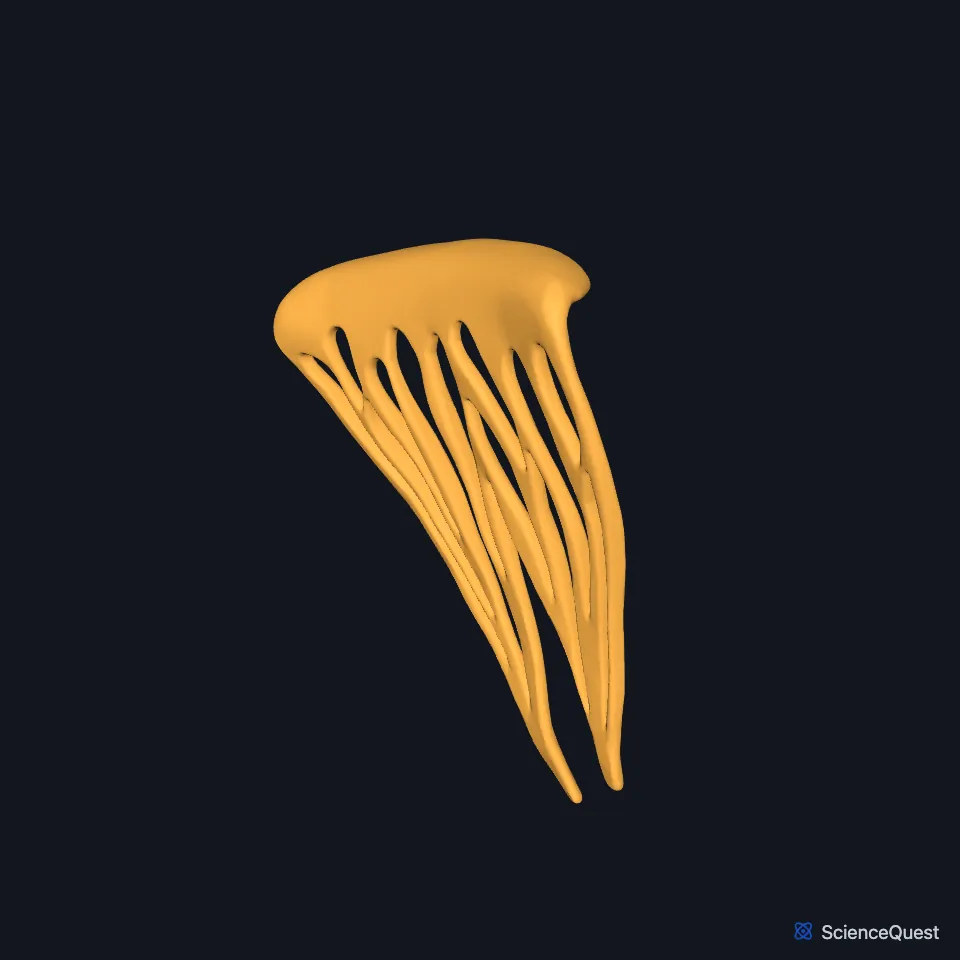
Where is the mitral valve?
It sits in the fibrous skeleton between the atria and the ventricles, where fibrous rings surround the four openings: two from the atria into the ventricles, and two from the ventricles into the pulmonary trunk and the aorta. Those rings anchor the valve leaflets. (OpenStax A&P 2e, 19.1; Gray 1918, 4b. The Heart; StatPearls, Histology, Heart)
Its anterior leaflet lies against the back of the aortic root and is fixed to it. (StatPearls, Mitral Valve)
On the chest wall, its closing is loudest in the fifth left intercostal space at the midclavicular line. (StatPearls, Heart Sounds)
What are the parts of the mitral valve?
The valve works as one apparatus: the mitral annulus, an anterior and a posterior leaflet, the chordae tendineae and the papillary muscles. (StatPearls, Mitral Valve)
The annulus is a fibrous oval ring around the opening. It contracts during systole and narrows the opening as the leaflets close. (StatPearls, Mitral Valve)
The anterior leaflet is the larger and thicker of the two. It takes up about a third of the ring and two thirds of the opening, and the posterior leaflet takes up the other two thirds of the ring. (StatPearls, Mitral Valve)
Chordae tendineae tie both leaflets to two papillary muscles that rise from the wall of the left ventricle, and each muscle sends cords to both leaflets. (OpenStax A&P 2e, 19.1; StatPearls, Mitral Valve)
The two muscles have two pairs of names: anterior and posterior, or anterolateral and posteromedial. OpenStax uses the first pair, the StatPearls article on the papillary muscles the second, and the StatPearls article on the mitral valve both. The longer names come from where the muscles sit in relation to the mitral commissures. (OpenStax A&P 2e, 19.1; StatPearls, Heart Papillary Muscles and Mitral Valve)
What does the mitral valve do?
While the heart relaxes, the valve stands open and blood flows from the left atrium into the left ventricle. Toward the end of that relaxation the left atrium contracts and pushes more blood in. (OpenStax A&P 2e, 19.3 and 19.1; StatPearls, Cardiac Cycle and Heart; Gray 1918, 4b. The Heart)
When the left ventricle starts to contract, its rising pressure closes the valve, marking the start of systole. (OpenStax A&P 2e, 19.3; StatPearls, Cardiac Cycle; Gray 1918, 4b. The Heart)
The papillary muscles contract with the ventricle and pull the chordae tight, which holds the cusps in place and stops them turning inside out into the atrium. (OpenStax A&P 2e, 19.1; Gray 1918, 4b. The Heart)
Its closing, with the tricuspid valve’s, makes the first heart sound, S1. The mitral part is the louder and comes first, because the left ventricle contracts earlier. (StatPearls, Heart Sounds)
What does the mitral valve connect to?
The left atrium above it receives blood from the lungs through four pulmonary veins. The left ventricle below it sends that blood through the aortic valve into the aorta. (OpenStax A&P 2e, 19.1; StatPearls, Heart)
The anterior papillary muscle draws blood from the anterior interventricular and circumflex arteries. The posterior one most often draws from the right coronary artery, and in some hearts from the circumflex. (StatPearls, Mitral Valve and Heart Papillary Muscles)
What can go wrong with the mitral valve?
Mitral stenosis, a narrowed valve, most often follows rheumatic heart disease. Fused papillary muscles can also narrow it, and so can a parachute mitral valve, in which all the chordae attach to a single papillary muscle. (StatPearls, Mitral Valve and Heart Papillary Muscles)
If a papillary muscle ruptures, which can happen after a myocardial infarction or a traumatic injury, the valve leaks: blood flows back into the left atrium (mitral regurgitation). (StatPearls, Heart Papillary Muscles and Mitral Valve; OpenStax A&P 2e, 19.1)
Where the sources differ
The sources rank mitral regurgitation differently. OpenStax calls it the most common valve disorder, found in about 2 percent of people. The StatPearls article on the mitral valve ranks it second, after aortic stenosis, and puts it at about 2 million people in the United States. (OpenStax A&P 2e, 19.1; StatPearls, Mitral Valve)
| Feature | Detail and source |
|---|---|
| Between | Left atrium and left ventricle (OpenStax A&P 2e, 19.1; StatPearls, Histology, Heart) |
| Cusps | Two: anterior and posterior (StatPearls, Mitral Valve) |
| Other names | Bicuspid valve, left atrioventricular valve (OpenStax A&P 2e, 19.1; FMA, FMA7235; Gray 1918, 4b. The Heart) |
| Papillary muscles | Two, joined to the cusps by chordae tendineae (OpenStax A&P 2e, 19.1; Gray 1918, 4b. The Heart) |
| Closes | As the ventricles contract, making the first heart sound (S1) (OpenStax A&P 2e, 19.3; StatPearls, Heart Sounds) |
Touches in this model
Measured on the BodyParts3D meshes: surfaces within 2 mm of the mitral valve, nearest first.
- Posterior papillary muscle of left ventricle in contact
- Wall of heart in contact
Sources
- OpenStax Anatomy and Physiology 2e, section 19.1, Heart Anatomy. CC BY-NC-SA 4.0. Read 2026-10-25.
- Gray’s Anatomy of the Human Body, 20th US edition (1918), V. Angiology, 4b. The Heart (1918 terms: longitudinal sulci for interventricular sulci, musculi papillares for papillary muscles). Henry Gray. Public domain. Read 2026-10-25.
- Foundational Model of Anatomy, via the EMBL-EBI Ontology Lookup Service, Mitral valve (FMA7235), preferred name and synonyms. Not printed on the page. Read 2026-10-25.
- StatPearls, Histology, Heart. Anita Arackal; Khalid Alsayouri. Last update January 2, 2023. CC BY-NC-ND 4.0. Read 2026-10-25.
- StatPearls, Anatomy, Thorax, Mitral Valve. Felipe Sanchez Vaca; Bruno Bordoni. Last update July 24, 2023. CC BY-NC-ND 4.0. Read 2026-10-25.
- StatPearls, Physiology, Heart Sounds. Sean Dornbush; Andre E. Turnquest. Last update July 17, 2023. CC BY-NC-ND 4.0. Read 2026-10-25.
- StatPearls, Anatomy, Thorax, Heart Papillary Muscles. Natalie L. Rich; Yusuf S. Khan. Last update July 24, 2023. CC BY-NC-ND 4.0. Read 2026-10-25.
- OpenStax Anatomy and Physiology 2e, section 19.3, Cardiac Cycle. CC BY-NC-SA 4.0. Read 2026-10-25.
- StatPearls, Physiology, Cardiac Cycle. Joshua D. Pollock; Amgad N. Makaryus. Last update October 3, 2022. CC BY-NC-ND 4.0. Read 2026-10-25.
- StatPearls, Anatomy, Thorax, Heart. Ibraheem Rehman; Afzal Rehman. Last update August 28, 2023. CC BY-NC-ND 4.0. Read 2026-10-25.
The values are facts and free to use; this page’s selection and presentation are © 2026 ScienceQuest.