Spleen
The spleen is the largest organ of the lymphatic system. It filters the blood and mounts immune responses to pathogens carried in it. (StatPearls, Spleen; OpenStax A&P 2e, 21.1)
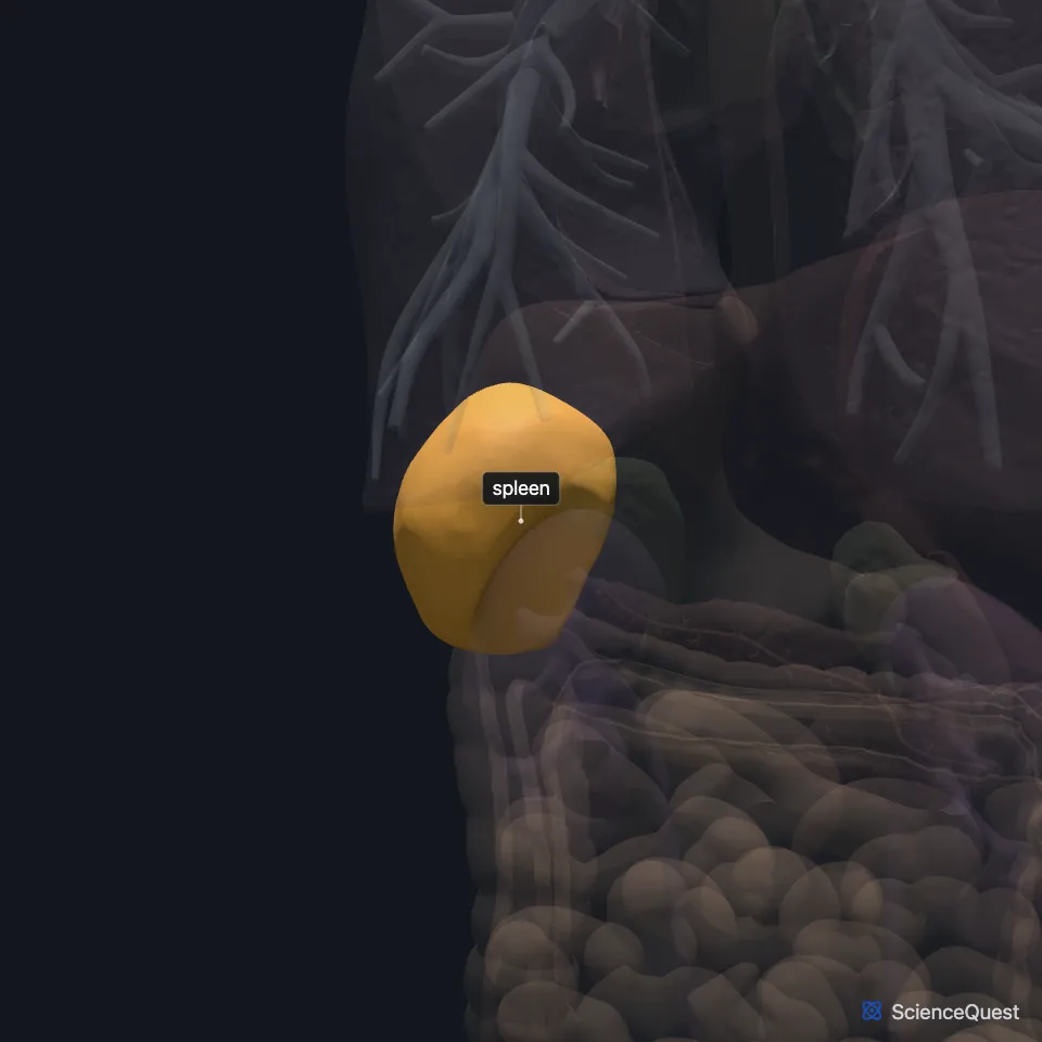
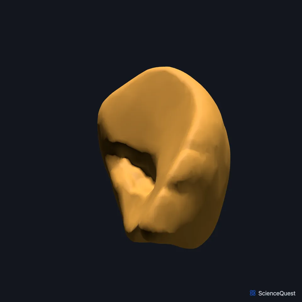
Where is the spleen?
It lies in the left hypochondriac region, between the fundus of the stomach and the diaphragm, level with the ninth to eleventh ribs. It cannot normally be felt on examination. (StatPearls, Spleen)
The gastrosplenic ligament ties it to the greater curvature of the stomach. (OpenStax A&P 2e, 21.1; StatPearls, Spleen; Gray’s Anatomy (1918), The Stomach)
It is one of the organs behind the stomach, on the far side of the lesser sac, along with the pancreas and the left kidney. (StatPearls, Stomach)
What is the spleen made of?
It measures about 10 to 12 cm and weighs about 150 to 200 g. A thin connective tissue capsule encloses it, and its many blood vessels make it dark red. (StatPearls, Spleen; OpenStax A&P 2e, 21.1)
Red pulp, mostly red blood cells, makes up most of its tissue. White pulp, which resembles the lymphoid follicles of lymph nodes, sits around central arterioles, and a marginal zone separates the two. (OpenStax A&P 2e, 21.1; StatPearls, Spleen)
What does the spleen do?
Red pulp filters the blood and removes old or damaged red blood cells. Its macrophages destroy viruses, bacteria and fungi. (StatPearls, Spleen)
White pulp is where T and B cell responses are mounted. The marginal zone steers pathogens from the blood into the white pulp. (OpenStax A&P 2e, 21.1; StatPearls, Spleen)
Red pulp also keeps a reserve of white cells and platelets that can be sent out after injury or blood loss. (StatPearls, Spleen)
Macrophages in its red pulp break down old red cells and recycle their iron. The splenic vein drains it into the portal vein, which leads to the liver. (OpenStax A&P 2e, 23.6; StatPearls, Physiology, Spleen)
Before birth the liver and spleen help make blood cells until the bone marrow takes over. (OpenStax A&P 2e, 21.1; StatPearls, Physiology, Spleen and Spleen)
It is not a vital organ, because red bone marrow, the liver and the lymph nodes can make up for its work. (StatPearls, Spleen)
What touches and supplies the spleen?
Its convex diaphragmatic surface is smooth. Its concave visceral surface carries impressions of the stomach, the left kidney, the colon and the tail of the pancreas. (StatPearls, Spleen)
The colon turns down below it at the left colic (splenic) flexure. The phrenicocolic ligament runs from that flexure to the diaphragm and supports the spleen from below. (OpenStax A&P 2e, 23.5; Gray’s Anatomy (1918), The Peritoneum; StatPearls, Large Intestine)
The splenic artery, from the celiac trunk, runs along the top of the pancreas to the hilum. Near the spleen it splits into 5 branches that do not join, so the organ has separate vascular segments. (StatPearls, Spleen)
The splenic vein leaves the hilum, passes behind the pancreas and joins the superior mesenteric vein to form the portal vein. (StatPearls, Spleen)
Its hilum also anchors the splenorenal and gastrosplenic ligaments. Sympathetic nerves come from the celiac plexus and parasympathetic nerves from the vagus. (StatPearls, Spleen)
Why is the spleen often injured?
It is among the abdominal organs injured most often, and a fragment of a broken left rib can puncture it. Some people also have accessory spleens, small pieces of splenic tissue apart from the main organ. (StatPearls, Spleen)
Where the sources differ
StatPearls chapters differ on the spleen’s share of fetal blood making. One says it becomes the major site of red cell production from week 12 until week 28, though the same chapter also calls it only partly responsible from week 10 to week 25; another says the liver is the dominant site and the spleen contributes only briefly, around week 20. (StatPearls, Physiology, Spleen and Embryology, Hematopoiesis)
| Feature | Detail and source |
|---|---|
| Size | About 10 to 12 cm; 150 to 200 g (StatPearls, Spleen) |
| Position | Left hypochondriac region, ninth to eleventh ribs (StatPearls, Spleen) |
| Tissue | Red pulp and white pulp, with a marginal zone between them (StatPearls, Spleen) |
| Artery | Splenic artery, from the celiac trunk (StatPearls, Spleen) |
| Vein | Splenic vein, joining the superior mesenteric vein to form the portal vein (StatPearls, Spleen) |
| Nerves | Celiac plexus (sympathetic) and vagus nerve (parasympathetic) (StatPearls, Spleen) |
Touches in this model
Measured on the BodyParts3D meshes: surfaces within 2 mm of the spleen, nearest first.
- Colon in contact
- Left kidney in contact
- Omental taenia in contact
- Stomach in contact
- Diaphragm in contact
- Splenic artery in contact
- Splenic vein in contact
- Left transversus abdominis 0.2 mm away
- Lower lobe of left lung 0.3 mm away
- Skin 0.3 mm away
- Left adrenal gland 1.9 mm away
Sources
- StatPearls, Anatomy, Abdomen and Pelvis, Spleen. Adekunle E. Omole; Marjorie V. Launico. Last update January 31, 2026. CC BY-NC-ND 4.0. Read 2026-10-25.
- OpenStax Anatomy and Physiology 2e, section 21.1, Anatomy of the Lymphatic and Immune Systems. CC BY-NC-SA 4.0. Read 2026-10-25.
- Gray’s Anatomy of the Human Body, 20th US edition (1918), Splanchnology, The Stomach, page 1162. Henry Gray; edited by Warren H. Lewis. Public domain. Read 2026-10-25.
- StatPearls, Anatomy, Abdomen and Pelvis: Stomach. Shazia R. Chaudhry; Maria Nataly P. Liman; Adekunle E. Omole; Diana C. Peterson. Last update July 17, 2024. CC BY-NC-ND 4.0. Read 2026-10-25.
- OpenStax Anatomy and Physiology 2e, section 23.6, Accessory Organs in Digestion: The Liver, Pancreas, and Gallbladder. CC BY-NC-SA 4.0. Read 2026-10-25.
- StatPearls, Physiology, Spleen. Vaishali Kapila; Chase J. Wehrle; Faiz Tuma. Last update May 1, 2023. CC BY-NC-ND 4.0. Read 2026-10-25.
- OpenStax Anatomy and Physiology 2e, section 23.5, The Small and Large Intestines. CC BY-NC-SA 4.0. Read 2026-10-25.
- Gray’s Anatomy of the Human Body, 20th US edition (1918), Splanchnology, The Peritoneum, page 1158. Henry Gray; edited by Warren H. Lewis. Public domain. Read 2026-10-25.
- StatPearls, Anatomy, Abdomen and Pelvis: Large Intestine. Adekunle E. Omole; Pujyitha Mandiga; Preet Kahai; Stany Lobo. Last update April 6, 2025. CC BY-NC-ND 4.0. Read 2026-10-25.
- StatPearls, Embryology, Hematopoiesis. Ranbir Singh; Kavin Sugumar. Last update May 3, 2025. CC BY-NC-ND 4.0. Read 2026-10-25.
The values are facts and free to use; this page’s selection and presentation are © 2026 ScienceQuest.