Jejunum
The jejunum is the middle part of the small intestine, running from the duodenum to the ileum. Its name is Latin for “empty”. (OpenStax A&P 2e, 23.5; StatPearls, Small Intestine)
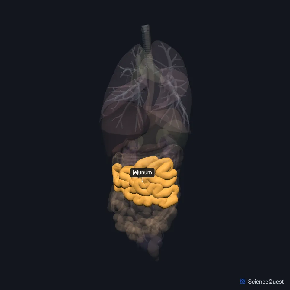
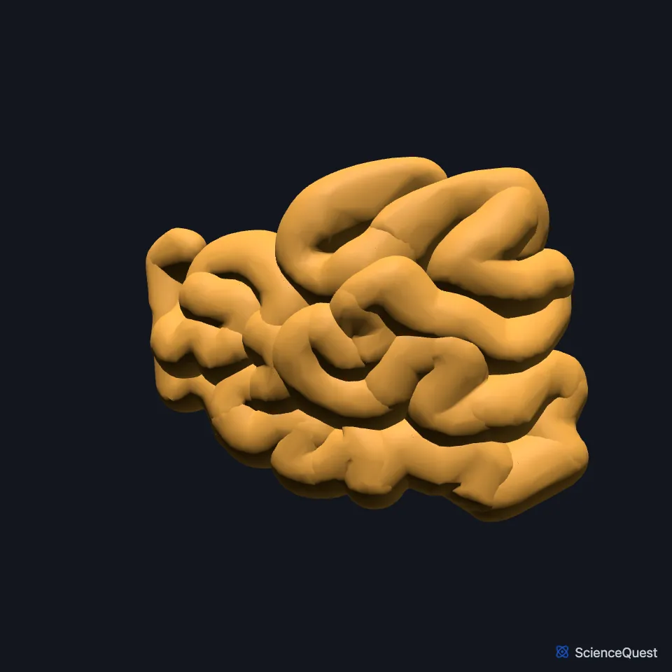
Where is the jejunum?
It begins at the duodenojejunal junction, where the gut comes back inside the peritoneum. The mesentery, a double fold of peritoneum, tethers it to the posterior abdominal wall. (StatPearls, Small Intestine; OpenStax A&P 2e, 23.5)
The large intestine frames the jejunum and the rest of the small intestine. (OpenStax A&P 2e, 23.5; StatPearls, Physiology, Small Bowel)
How is the jejunum built for absorption?
Its lining carries circular folds (plicae circulares) and villi. The folds make chyme spiral through instead of moving straight, which slows it and gives time to absorb nutrients. (StatPearls, Small Intestine; OpenStax A&P 2e, 23.5)
Circular folds and villi enlarge its absorptive surface, and each enterocyte carries about 3,000 microvilli. (OpenStax A&P 2e, 23.5; StatPearls, Physiology, Small Bowel)
Each villus holds a capillary bed and a lymph capillary called a lacteal. Amino acids and most other nutrients enter the blood in the capillaries, while lipids enter the lacteals and travel in lymph to the thoracic duct. (OpenStax A&P 2e, 23.5; StatPearls, Physiology, Nutrient Absorption)
What does the jejunum do?
It absorbs mainly carbohydrates, amino acids and fatty acids through its villi. (StatPearls, Small Intestine)
With the duodenum, its upper part is the main site where dietary iron is absorbed. (StatPearls, Small Intestine)
Between meals, migrating motor complexes sweep undigested material along the stomach and small intestine toward the large intestine. (OpenStax A&P 2e, 23.5; StatPearls, Physiology, Motilin)
Endocrine cells in the upper small intestine, which StatPearls calls Mo cells, release motilin in cycles during fasting. Motilin stimulates movement in the stomach and small intestine. (OpenStax A&P 2e, 23.5; StatPearls, Physiology, Motilin)
What supplies the jejunum?
Jejunal arteries from the superior mesenteric artery run through the mesentery. They form arterial arcades, which give off the vasa recta to the gut wall. (StatPearls, Small Intestine)
Its veins join the superior mesenteric vein, which meets the splenic vein to form the portal vein. That carries the nutrient-rich blood to the liver. (StatPearls, Small Intestine; OpenStax A&P 2e, 23.5)
Vagal fibers increase its secretion and movement. Sympathetic fibers reach it through the superior mesenteric plexus and do the opposite, also narrowing its vessels. (StatPearls, Small Intestine)
Where the sources differ
The sources disagree on how to tell the jejunum from the ileum. OpenStax says the ileum is thicker, more vascular and has more developed mucosal folds; StatPearls gives the jejunum the thicker wall, the greater vascularity and a deeper red color. (OpenStax A&P 2e, 23.5; StatPearls, Small Intestine)
Sources differ on how much the lining enlarges the absorptive surface. StatPearls puts the lining of the duodenum and jejunum at about 100 times the area of the skin, and OpenStax says folds, villi and microvilli enlarge the small intestine’s surface more than 600-fold. A 2014 review that measured biopsies found the folds add about 1.6 times and villi with microvilli 60 to 120 times, and put the whole digestive tract lining at about 32 square meters, half a badminton court rather than the tennis court textbooks give. (StatPearls, Physiology, Small Bowel; OpenStax A&P 2e, 23.5; Helander and Fändriks 2014, Scand J Gastroenterol)
They also give very different lengths: about 0.9 m (3 ft) in life in OpenStax, roughly 2.5 m in StatPearls. (OpenStax A&P 2e, 23.5; StatPearls, Small Intestine)
| Feature | Detail and source |
|---|---|
| Runs | From the duodenojejunal junction to the ileum, with no clear boundary (StatPearls, Small Intestine; OpenStax A&P 2e, 23.5) |
| Held by | The mesentery, to the posterior abdominal wall (OpenStax A&P 2e, 23.5; StatPearls, Small Intestine) |
| Lining | Plicae circulares and villi (StatPearls, Small Intestine) |
| Absorbs | Mainly carbohydrates, amino acids and fatty acids (StatPearls, Small Intestine) |
| Arteries | Jejunal arteries from the superior mesenteric artery (StatPearls, Small Intestine) |
Touches in this model
Measured on the BodyParts3D meshes: surfaces within 2 mm of the jejunum, nearest first, 12 of 14.
- Colon in contact
- Duodenum in contact
- Free taenia in contact
- Ileum in contact
- Pancreas in contact
- Left internal oblique in contact
- Left rectus abdominis in contact
- Left transversus abdominis in contact
- Linea alba in contact
- Right internal oblique in contact
- Right transversus abdominis in contact
- Skin in contact
Sources
- OpenStax Anatomy and Physiology 2e, section 23.5, The Small and Large Intestines. CC BY-NC-SA 4.0. Read 2026-10-25.
- StatPearls, Anatomy, Abdomen and Pelvis, Small Intestine. Jason T. Collins; Amanda Nguyen; Adekunle E. Omole; Madhu Badireddy. Last update February 18, 2025. CC BY-NC-ND 4.0. Read 2026-10-25.
- StatPearls, Physiology, Small Bowel. Elizabeth M. Fish; Karlie R. Shumway; Bracken Burns. Last update January 31, 2024. CC BY-NC-ND 4.0. Read 2026-10-25.
- StatPearls, Physiology, Nutrient Absorption. Eric J. Basile; Krunal Shukla; Marjorie V. Launico; Amy J. Sheer. Last update December 1, 2025. CC BY-NC-ND 4.0. Read 2026-10-25.
- StatPearls, Physiology, Motilin. Murtaja Z. Al-Missri; Ishwarlal Jialal. Last update September 26, 2022. CC BY-NC-ND 4.0. Read 2026-10-25.
- Scandinavian Journal of Gastroenterology, Surface area of the digestive tract, revisited, Scand J Gastroenterol 49(6), pages 681 to 689 (abstract, PMID 24694282). Helander HF, Fändriks L. Abstract read on Europe PMC; no reuse licence stated. Read 2026-10-25.
The values are facts and free to use; this page’s selection and presentation are © 2026 ScienceQuest.