Superior mesenteric artery
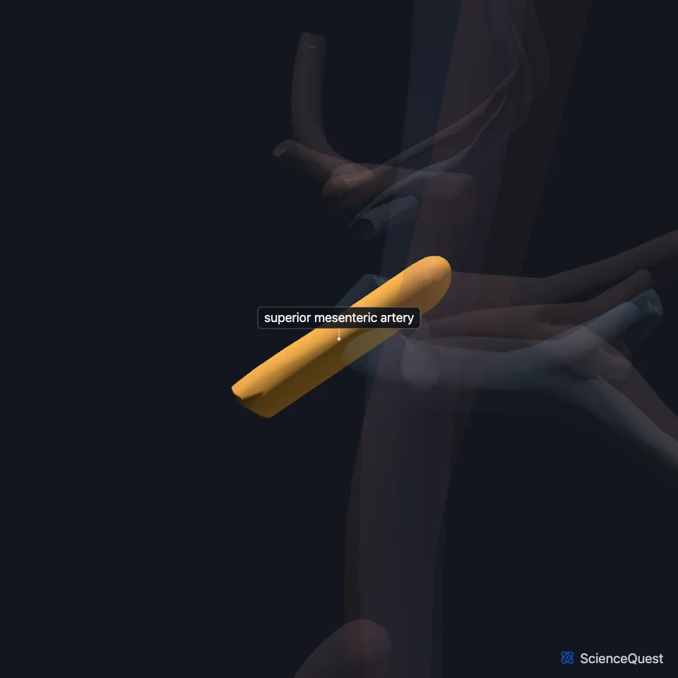
The superior mesenteric artery is the second major branch of the abdominal aorta. It supplies the small intestine from the second part of the duodenum, the head of the pancreas, and the large intestine as far as the splenic flexure. (StatPearls, Abdomen and Pelvis: Superior Mesenteric Artery; OpenStax A&P 2e, 20.5)
Where is the superior mesenteric artery?
It leaves the front of the aorta at the level of L1, below the celiac trunk and above the renal arteries. (StatPearls, Abdomen and Pelvis: Superior Mesenteric Artery)
What does the superior mesenteric artery supply?
It supplies the third part of the duodenum, the jejunum, the ileum, the cecum, the ascending colon and the proximal transverse colon. (StatPearls, Abdomen and Pelvis: Superior Mesenteric Artery)
Its inferior pancreaticoduodenal branch, with the superior pancreaticoduodenal artery, supplies the head of the pancreas. (StatPearls, Abdomen and Pelvis: Superior Mesenteric Artery)
Its right colic, ileocolic and middle colic branches supply the colon. The marginal artery of Drummond links them with the inferior mesenteric branches along the mesenteric border. (StatPearls, Abdomen and Pelvis: Superior Mesenteric Artery)
What lies in front of and behind the superior mesenteric artery?
In front lie the pylorus of the stomach, the neck of the pancreas and the splenic vein. Behind lie the uncinate process of the pancreas, the lower duodenum and the left renal vein. (StatPearls, Abdomen and Pelvis: Superior Mesenteric Artery)
The left renal vein passes between the aorta and the origin of the superior mesenteric artery. (StatPearls, Abdomen and Pelvis: Superior Mesenteric Artery)
The superior mesenteric vein joins the splenic vein at about L2 to form the hepatic portal vein. (OpenStax A&P 2e, 20.5; Gray’s Anatomy (1918), The Portal System of Veins; StatPearls, Abdomen and Pelvis: Superior Mesenteric Vein)
How did the superior mesenteric artery form?
It grows from one of the paired ventral segmental arteries of the embryo’s dorsal aorta. The 10th, 13th and 21st persist as the celiac trunk and the superior and inferior mesenteric arteries. (StatPearls, Abdomen and Pelvis: Celiac Trunk)
Where the sources differ
The sources give different gaps between it and the celiac trunk: about 2.5 cm in OpenStax, about 1 cm in StatPearls. (OpenStax A&P 2e, 20.5; StatPearls, Abdomen and Pelvis: Superior Mesenteric Artery)
| Feature | Detail and source |
|---|---|
| Arises from | Front of the abdominal aorta, at L1 (StatPearls, Abdomen and Pelvis: Superior Mesenteric Artery) |
| Supplies | Small intestine, head of the pancreas, large intestine as far as the splenic flexure (OpenStax A&P 2e, 20.5; StatPearls, Abdomen and Pelvis: Superior Mesenteric Artery) |
| Colic branches | Right colic, ileocolic, middle colic (StatPearls, Abdomen and Pelvis: Superior Mesenteric Artery) |
Touches in this model
Measured on the BodyParts3D meshes: surfaces within 2 mm of the superior mesenteric artery, nearest first.
- Descending aorta in contact
- Left renal artery in contact
- Left renal vein in contact
- Pancreas in contact
- Superior mesenteric vein 0.2 mm away
- Right renal artery 1.4 mm away
Sources
- StatPearls, Anatomy, Abdomen and Pelvis: Superior Mesenteric Artery. Adekunle E. Omole; Arshia Khorasani-Zadeh. Last update September 14, 2026. CC BY-NC-ND 4.0. Read 2026-10-25.
- OpenStax Anatomy and Physiology 2e, section 20.5, Circulatory Pathways. CC BY-NC-SA 4.0. Read 2026-10-25.
- Anatomy of the Human Body (Gray’s Anatomy), 20th US edition, 1918, Angiology, The Portal System of Veins, the portal vein; Internet Archive scan of the 1918 Lea and Febiger printing, terminology of 1918. Henry Gray; edited by Warren H. Lewis. Public domain. Read 2026-10-25.
- StatPearls, Anatomy, Abdomen and Pelvis: Superior Mesenteric Vein. Andrew Broussard; Chase J. Wehrle; Navdeep S. Samra. Last update August 8, 2023. CC BY-NC-ND 4.0. Read 2026-10-25.
- StatPearls, Anatomy, Abdomen and Pelvis: Celiac Trunk. Dr. Rohin Garg; Ali Nassereddin; Tafline C. Arbor. Last update September 15, 2025. CC BY-NC-ND 4.0. Read 2026-10-25.
The values are facts and free to use; this page’s selection and presentation are © 2026 ScienceQuest.