Superior mesenteric vein
The superior mesenteric vein collects most of the venous blood from the jejunum and ileum. Behind the neck of the pancreas it joins the splenic vein to form the portal vein. (StatPearls, Abdomen and Pelvis: Superior Mesenteric Vein; Gray’s Anatomy (1918), The Portal System of Veins)
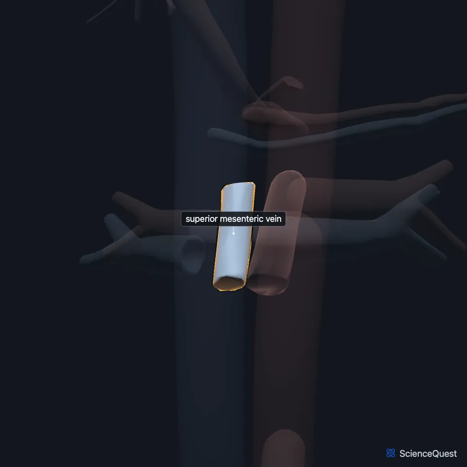
Where is the superior mesenteric vein?
It begins in the right iliac fossa, from the veins of the end of the ileum, the cecum and the appendix, and rises between the two layers of the mesentery. (Gray’s Anatomy (1918), The Portal System of Veins)
On the way up it passes in front of the right ureter, the inferior vena cava, the inferior part of the duodenum and the lower part of the head of the pancreas. (Gray’s Anatomy (1918), The Portal System of Veins)
What does the superior mesenteric vein drain?
Its tributaries match the branches of the superior mesenteric artery: the intestinal, ileocolic, right colic and middle colic veins. It also takes the right gastroepiploic vein from the stomach and the pancreaticoduodenal veins. (Gray’s Anatomy (1918), The Portal System of Veins; StatPearls, Abdomen and Pelvis: Superior Mesenteric Vein)
Through the portal vein its blood passes through the liver. The portal venous system supplies about 75% of the liver’s blood. (StatPearls, Abdomen and Pelvis: Superior Mesenteric Vein and Abdomen and Pelvis, Portal Venous System)
What lies next to the superior mesenteric vein?
It normally stays on the right of the superior mesenteric artery. A vein on the left of the artery can mean midgut malrotation or a tumor pressing on it, though it can also be a passing finding in a normally rotated gut. (StatPearls, Abdomen and Pelvis: Superior Mesenteric Vein)
How does the superior mesenteric vein vary?
It may be one trunk, 5 to 50 mm long, before it splits into right and left intestinal branches. A common variant has two trunks that drain straight into the junction with the splenic vein. (StatPearls, Abdomen and Pelvis: Superior Mesenteric Vein)
How does the superior mesenteric vein develop?
Older accounts treat it as what remains of the left vitelline vein. A 2017 study of 5 to 6 week embryos suggests instead that it forms within the mesentery as that vein regresses. (StatPearls, Abdomen and Pelvis: Superior Mesenteric Vein)
What is superior mesenteric vein thrombosis?
A clot limited to this vein is reported to run a milder course than one that also fills the portal vein, because its many collateral veins protect the bowel from infarction. Computed tomography is the gold standard test. (StatPearls, Abdomen and Pelvis: Superior Mesenteric Vein)
Where the sources differ
The sources draw its colonic territory differently. StatPearls (Superior Mesenteric Vein) has it drain the large intestine up to the splenic flexure, StatPearls (Portal Venous System) up to the first two-thirds of the transverse colon, and Gray’s Anatomy (1918) names the cecum and the ascending and transverse colon. (StatPearls, Abdomen and Pelvis: Superior Mesenteric Vein and Abdomen and Pelvis, Portal Venous System; Gray’s Anatomy (1918), The Portal System of Veins)
| Feature | Detail and source |
|---|---|
| Begins | Right iliac fossa (Gray’s Anatomy (1918), The Portal System of Veins) |
| Ends | Joins the splenic vein behind the neck of the pancreas to form the portal vein (StatPearls, Abdomen and Pelvis: Superior Mesenteric Vein) |
| Lies | To the right of the superior mesenteric artery (StatPearls, Abdomen and Pelvis: Superior Mesenteric Vein) |
Touches in this model
Measured on the BodyParts3D meshes: surfaces within 2 mm of the superior mesenteric vein, nearest first.
- Inferior vena cava in contact
- Left renal vein in contact
- Pancreas in contact
- Superior mesenteric artery 0.2 mm away
Sources
Sources read
- StatPearls, Anatomy, Abdomen and Pelvis: Superior Mesenteric Vein. Andrew Broussard; Chase J. Wehrle; Navdeep S. Samra. Last update . CC BY-NC-ND 4.0. Read .
- Anatomy of the Human Body (Gray’s Anatomy), 20th US edition, 1918, Angiology, The Portal System of Veins, the portal vein; Internet Archive scan of the 1918 Lea and Febiger printing, terminology of 1918. Henry Gray; edited by Warren H. Lewis. Public domain. Read .
- StatPearls, Anatomy, Abdomen and Pelvis, Portal Venous System (Hepatic Portal System). Joshua M. Harkins; Bareeqah Ahmad. Last update . CC BY-NC-ND 4.0. Read .
The values are facts and free to use; this page’s selection and presentation are © 2026 ScienceQuest.